|
Corresponding author: Alexandre Loukanov ( alex_loukanov@yahoo.com ) Academic editor: Georgi Momekov
© 2019 Alexandre Loukanov, Svetla Nikolova, Chavdar Filipov, Seiichiro Nakabayashi.
This is an open access article distributed under the terms of the Creative Commons Attribution License (CC BY 4.0), which permits unrestricted use, distribution, and reproduction in any medium, provided the original author and source are credited.
Citation:
Loukanov A, Nikolova S, Filipov C, Nakabayashi S (2019) Nanomaterials for cancer medication: from individual nanoparticles toward nanomachines and nanorobots. Pharmacia 66(3): 147-156. https://doi.org/10.3897/pharmacia.66.e37739
|
Abstract
The nanomaterials for cancer medication are already reality providing a wide range of new tools and possibilities, from earlier diagnostics and improved imaging to better, more efficient, and more targeted anticancer therapies. The purpose of this critical review is to focus on the current use of clinically approved nanoparticles for cancer theranostic, nanovaccines and delivery platforms for gene therapy. These include inorganic, metal and polymer nanoparticles, nanocrystals and varieties of drug delivery nanosystems (micelles, liposomes, microcapsules and etc.). The recent progress in cancer nanomedicine enables to combine the benefits of individual nanoparticles with biomolecules into a multifunction nanomachines and even highly advanced nanorobots for targeted therapies. Nowadays clinical trials with advanced anticancer nanomachines provide potential for more accurately and effective identification and destruction of the cancer cells present in the human body.
Keywords
Drug delivery nanosystems, targeted nanotherapy, nanomachines, personalized nanomedicine
Introduction
The conventional approaches to combating cancer are limited to radiation, drug chemotherapy, and surgery, which are often not satisfactory treatments of patients with metastatic cancer. They simultaneously damage numerous healthy tissues and thus induce a lot of harmful side effects that accompanies these treatments. The nanoparticles have potential to be used as an alternative medication since they offer great benefits for targeted drug delivery directly to cancerous cells and neoplasms and enhance the therapeutic efficacy (

Nanocrystals of drug molecules are approved for oral, local and intravenous administration because of their high drug-loading efficiency, great structural stability, long circulation time and steady dissolution (
Liposome systems for drugs encapsulating are used in some cancer therapies, because of the benefit to provide targeting of the anticancer substance, respectively in lower dose, which cause significantly reduced toxicity on the normal cells (

Lipid nanocapsules (LNC) with size distribution 25~110 nm have ability to encapsulate efficiently lipophilic drugs (etoposide, docetaxel, hydroxytamoxifen, paclitaxel, etc.), offering a pharmaceutical solution for their intravenous administration (
Polymer nanoparticles provide sustained and time-dependent release of drugs (paclitaxel, gemcitabine, anthracycline, irinotecan, etc.), thereby controlled therapeutic approach (
The presented above overview examined the various type nanomaterials for cancer medication. Below is discussed their clinical applications, which are already approved in the oncological practice.
Cancer nanotheranostics
The integration of diagnostic imaging and therapeutic functions in the same nanoparticles is highly beneficial for the personalized and precision nanomedicine. The gold nanoparticles (Au NPs) for cancer theranostics provide labeling, delivering, heating, sensing and detection (

The prostate cancer cells show enhanced inhibition if are simultaneously treated with glucose-bound gold nanoparticles and irradiation (
The gold/mesoporous silica hybrid nanoparticles express the advantages of both mesoporous silica nanoparticle and conventional gold nanoparticles, namely great drug loading capacity, perfect photothermal converting ability and controllable drug release (

(A) Schematic illustration on the fabrication and (B) electron microscopic image of doxorubicin@gold/mesoporous silica nanoparticles. The nanoparticles are prepared in dual surfactant system using classic fast self-assembling method containing both cationic and non-ionic surfactants. Scale bar: 50 nm.
The magnetic (iron oxide, Fe2O3 or Fe3O4) nanoparticles are used in cancer theranostics alone or loaded into polymer or liposomes as drug delivery platforms. They have prominent transverse relaxation time and are used successful as contrast materials for magnetic resonance imaging (MRI) or as hyperthermia agent, drug and gene delivery carriers (feridex, endorem, gastomark, sinerem). Iron oxide nanoparticles might produce reactive oxygen species by Fenton reaction and thus disturb the cellular homeostasis, alter the intracellular signaling and pathways of cancer functioning. The liposomes and micelles platforms can also encapsulate fluorescent dyes for optical detection or radionuclides for positron emission tomography (PET) imaging.
The nanoparticles of polyethylene glycol (PEG), poly(D,L-lactic acid), poly(D,L-glycolic acid) and poly(ε-caprolactone) have already been approved as polymer-based platforms for clinical usage (e.g. oncaspar, renegal, neulasta, etc.). In general, the polymer nanoparticles offers stabilization and biocompatibility, contain a therapeutic agents (siRNA or small-molecule drug) as well as imaging agents (radionuclide, photosensitizer or MRI contrast agent as gadolinium or superparamagnetic iron oxide). Chlorin e6 or porphyrins are commonly used as photosensitizer agents in the laser therapy. The nanoparticles might be conjugated with targeting ligands to enhance the delivery in tumor site (
Cancer nanovaccines
The nanovaccines are designed to deliver antigens and/or adjuvants to tailor immune responses by specific targeting of immune cells (mostly dendritic cells or T cells). The cancer nanovaccination involves priming of the immune system with aim to induce a T-cell response against tumor-associated antigens. Nanoparticles-based cancer vaccines are already used to treat a wide array of cancers as melanoma (liposomes, carbon nanotubes), breast cancer (polymeric PEG nanoparticles functionalized with poly-lactic-co-glycolic acid or PLGA), prostate cancer (virus-like particles), etc. The polymeric nanoparticles are usually fabricated from biodegradable organics as PLGA, PEG, polycaprolactone, chitosan and dextran (
The vaccine liposomes are used as delivery systems for siRNA, DNA and hydrophilic or lipophilic antigens. The positively charged (cationic) liposomes induce strong immune response and are more efficiently taken by APCs-like macrophages and dendritic cells. For example, cationic DNA vaccine liposomes stimulate the long-lasting immune response for melanoma. OVA-loaded galactosylated liposomes demonstrated cytotoxic T lymphocytes (CTL) responses and remarkable antibody production against tumor growth (
The inorganic particles (gold, quantum dots, aluminium-based nanoparticles, iron oxide, etc.) are also viable delivery systems for cancer vaccines. The peptide-conjugated gold nanoparticles (Fig.

The magnetic nanoparticles (iron oxide) are also desirable potential vaccine systems for cancer therapy. There is significant differences in the immune response and respective tumor inhibition in case of OVA formulated with iron oxide, in comparison with only soluble OVA alone. The iron oxide nanoparticles greatly promote the activation of immune cells and cytokine production. Combination therapy with iron oxide cause a higher accumulation in lymph node dendritic cells and increase the immune response.
Biomimetic nanoparticles as high-density lipoprotein (HDL) can be uptaken without causing immunogenicity and toxicity. HDL-like nanoparticles act as drug-delivery systems and can incorporate inorganic nanoparticles, such as gold, iron oxide and quantum dots in design enables long-term circulation in the blood system with potential ability to vaccines or/and antigens. Such nanoparticles, which consist the fusion peptide α-Ap efficiently target dendritic cells through scavenger receptor class B1 (SR-B1)-mediated pathway.
The presented nanoparticles can be tuned through modulating of their size, shape, composition and surface properties to enhance the immune response against cancer. In clinical usage the therapeutic effect is achieved by enhanced immune response activation due to the nanoparticle-mediated cancer vaccines, which provide insight for prognosis and serve as a proper reference for the future nanoparticle research development.
Nano-platforms for tumor gene therapy
Synthetic nanoparticles (polymers and lipids) have been developed as platforms for nucleic acid delivery in the gene therapy of tumors and alternatives to the toxic and potentially oncogenes viral vectors. Their design might avoid the stimulation of immune system. The simplest drug delivery platforms are formed as polyplexes, i.e. electrostatic complexation between cationic polymers (e.g. mix of polythyleneimine, PEI and PEG) and plasmid DNA. The stealth liposomes are providing stable and safe alternatives too for effective delivery of nucleic acids, including siRNA and miRNA. DNA-liposomes, known as lipoplexes can be easily modified with targeting ligands with aim to enhance the site-specific delivery function. Cationic lipids and polymers can be combined with either DNA or RNA to form a stable nanoparticles, which are capable to gene transfer into the target cells. Such commercial drug (named JVRS-100) is used for anti-tumor responses generated by the immune system, because it can enters the macrophages and dendritic cells and activates Toll receptors (
The hyaluronic acid (HA) can form self-assembled organic nanoparticles for specifically delivery of siRNA (or other chemotherapeutic drugs) into malignant tumors. HA has negative charge which does not favor the complex formation with siRNA and due to this reason most nanocarriers include other cationic components (e.g. PEI), which influence the whole system’s gene silencing efficiency. The overexpressed receptors in cancerous cells for hyaluronic acid are known as CD44. Another more effective strategy to target CD44 receptors is the association of siRNA with both HA polyanion and chitosan polycation. Such delivery system is decisive for the successful application of siRNA or plasmid DNA-based death-induced gene therapy. Various challenges are still existing in the design of nano-platforms for perfect transfection vector, however the nanoparticles have been proved to be a suitable candidate.
Current development of anticancer nanomachines and nanorobots
Although numerous nanotherapies have received clinical approval, the most promising nanotechnology application for cancer medication still lie ahead. During the last decade the nanoparticles with anticancer activity and specificity were evolving in more complicate nanostructures, which mimic bioprocesses in the nature. Individual nanoparticles perform defined functions that may find application for some medical purposes, for example gold nanoparticles are utilized for treatment of the cancer of pancreas. However, a synergistic effect might be achieved if the benefits of various types’ nanoparticles are combined as in the case of hybrid gold/mesoporous nanoparticles, which have higher drug loading capacity and better photothermal converting ability then the single nanoparticles. However, the human organism possesses defense mechanisms, biological barriers and multiple strategies to protect against foreign xenobiotics or biological agents as bacteria, viruses and even nanoparticles. This challenge can be overcome by design of complicated nanomachines, which are capable of independently performing numerous tasks. The engineering and fabrication of nanomachines is innovative and promising field in the modern nanomedicine. They are designed to store, deliver, and release payload in response to intentionally caused stimuli (

Another example for design of light-powered nanomachine is the so called “nanoimpeller”. It is nanomechanical system that contains nano-sized mesoporous silica as a container to trap and release cargo molecules. The design allows of the nanoimpeller to be taken up by cancer cells in dark condition. The nanoimpeller releases its drug content from the tiny silica pores into the cancer cell interior when the specimen is irradiated (
In nanoscale design of nanorobots requires autonomous power supply, biosensors, nanomotors, etc. for overcoming of the low Reynolds number viscous drag and Brownian motion (
Conclusion and future directions
The medical nanotechnology opens new opportunities to improve significantly the cancer therapy by novel generation nanomaterials and nanomachines. The rapid progress in the development of nanomachine and nanorobotics technology doubtless will create powerful tools for effective treatment of various malignant tumors. The nanorobots used in medicine are predicted to provide a wealth of promise. It is possible in the near further, these tiny nanodevices to contribute the progress of advanced nano-medication for personalized medical care, and for establishment a new era in the screening, treatment, and prevention of cancer diseases.
References
- Allen C, Dos Santos N, Gallagher R, Chiu GNC, Shu Y, Li WM, Johnstone SA, Janoff AS, Mayer LD, Webb MS, Bally MB (2002) Controlling the physical behavior and biological performance of liposome formulations through use of surface grafted poly(ethylene glycol). Bioscience Reports 22(2): 225–250. https://doi.org/10.1023/A:1020186505848
- Anton N, Saulnier P, Gaillard C, Porcher E, Vrignaud S, Benoit JP (2009) Aqueous-core lipid nanocapsules for encapsulating fragile hydrophilic and/or lipophilic molecules. Langmuir 25(19): 11413–11419. https://doi.org/10.1021/la901565q
- Baratt G (2003) Colloidal drug carriers: Achievements and perspectives. Cellular and Molecular Life Sciences 60(1): 21–37. https://doi.org/10.1007/s000180300002
- Barratt GM (2000) Therapeutic applications of colloidal drug carriers. Pharmaceuticals Science and Technology Today 3(5): 163–171. https://doi.org/10.1016/S1461-5347(00)00255-8
- Bhattacharya R, Patra C, Earl A, Wang S, Katarya A, Lu L, Kizhakkedathu J, Yaszemski M, Greipp P, Mukhopadhyay D (2007) Attaching folic acid on gold nanoparticles using noncovalent interaction via different polyethylene glycol backbones and targeting of cancer cells. Nanomedicine: Nanotechnology, Biology and Medicine 3: 224–238. https://doi.org/10.1016/j.nano.2007.07.001
- Cattel L, Ceruti M, Dosio F (2003) From conventional to stealth liposomes: a new frontier in cancer chemotherapy. Tumori 89(3): 237–249. https://doi.org/10.1177/030089160308900302
- Chaubal MV (2003) Synthetic polymer-based ion-exchange resins: Excipients and activities. Drug Delivery Technology 3: 6–8.
- Cheng B, He H, Huang T, Berr SS, He J, Fan D, Zhang J, Xu P (2016) Gold Nanosphere Gated Mesoporous Silica Nanoparticle Responsive to Near-Infrared Light and Redox Potential as a Theranostic Platform for Cancer Therapy. Journal of Biomedical Nanotechnology 12(3): 435–449. https://doi.org/10.1166/jbn.2016.2195
- Chiellini E, Sunamoto J, Migliaresi C, Ottenbrite RM (2001) Biomedical Polymers and Polymer Therapeutics. Springer Verlag, Berlin. https://doi.org/10.1007/b112950
- Collins PG, Arnold MS, Avouris P (2001) Engineering carbon nanotubes and nanotube circuits using electrical breakdown. Science 2: 706–709. https://doi.org/10.1126/science.1058782
- Coon JS, Knudson W, Clodfelter K, Lu B, Weinstein RS (1991) Solutol HS 15, nontoxic polyoxyethylene esters of 12-hydroxystearic acid, reverses multidrug resistance. Cancer Research 51(3): 897–902.
- Dogra P, Butner JD, Chuang YI, Caserta S, Goel S, Brinker CJ, Cristini V, Wang Z (2019) Mathematical modeling in cancer nanomedicine: a review. Biomedical Microdevices 21: 1–40. https://doi.org/10.1007/s10544-019-0380-2
- Drexler KE, Merkle RC (2019) Simple Pump Selective for Neon. http://www.imm.org/Parts/Parts1.html [http://www.imm.org/Images/pumpApartC.jpg]
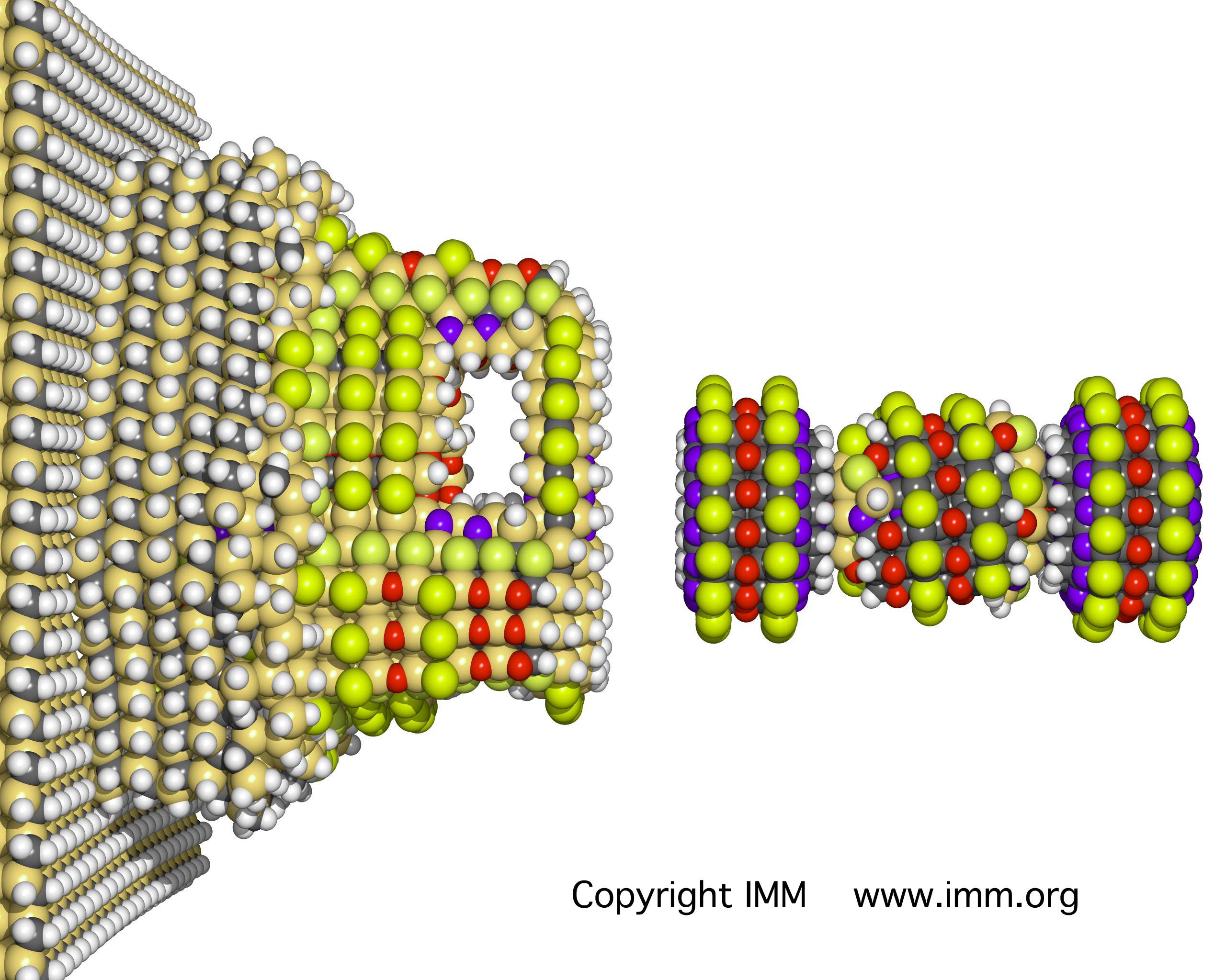
- Fang JY, Hwang TL, Huang YL (2006) Liposomes as vehicles for enhancing drug delivery via skin routes. Current Nanoscience 2(1): 55–70. https://doi.org/10.2174/157341306775473791
- Farokhzad OC, Jon S, Khademhosseini A, Tran TNT, LaVan DA, Langer R (2004) Nanoparticle-aptamer bioconjugates – A new approach for targeting prostate cancer cells. Cancer Research 64: 7668–7672. https://doi.org/10.1158/0008-5472.CAN-04-2550
- Felfoul O, Mohammadi M, Taherkhani S, de Lanauze D, Zhong XY, Loghin D, Essa S, Jancik S, Houle D, Lafleur M, Gaboury L, Tabrizian M, Kaou N, Atkin M, Vuong T, Batist G, Beauchemin N, Radzioch D, Martel S (2016) Magneto-aerotactic bacteria deliver drug-containing nanoliposomes to tumour hypoxic regions, Nature Nanotechnology 11: 941–947. https://doi.org/10.1038/nnano.2016.137
- Gao D, Agayan RR, Xu H, Philbert MA, Kopelman R (2006) Nanoparticles for two-photodynamic therapy in living cells. Nano Letters 6: 2383–2386. https://doi.org/10.1021/nl0617179
- Garcion E, Lamprecht A, Heurtault B, Paillard A, Aubert-Pouessel A, Denizot B, Menei P, Benoît JP (2006) A new generation of anticancer, drug-loaded, colloidal vectors reverses multidrug resistance in glioma and reduces tumor progression in rats. Molecular Cancer Therapeutics 5(7): 1710–1722. https://doi.org/10.1158/1535-7163.MCT-06-0289
- Gasco MR (1993) Method for producing solid lipid microspheres having a narrow size distribution. U.S. Patent 5: 250–236.
- Harisinghani MG, Barentsz J, Hahn PF, Deserno WM, Tabatabaei S, van de Kaa CH, de la Rosette J, Weissleder R (2003) Noninvasive detection of clinically occult lymph-node metastases in prostate cancer. New England Journal of Medicine 348: 2491–2499. https://doi.org/10.1056/NEJMoa022749
- Harris JM (1992) Poly(ethylene glycol) Chemistry: Biotechnological and Biomedical Applications (1st edn.). Springer, New York. https://doi.org/10.1007/978-1-4899-0703-5_1
- Heurtault B, Saulnier P, Pech B, Proust JE, Benoit JP (2002) A novel phase inversion-based process for the preparation of lipid nanocarriers. Pharmaceutical Research 19(6): 875–880. https://doi.org/10.1023/A:1016121319668
- Huynh NT, Passirani C, Saulnier P, Benoit JP (2009) Lipid nanocapsules: A new platform for nanomedicine. International Journal of Pharmaceutics 379(2): 201–209. https://doi.org/10.1016/j.ijpharm.2009.04.026
- Jiang P-L, Lin H-J, Wang H-W, Tsai W-Y, Lin S-F, Chien M-Y, Liang P-H, Huang Y-Y, Liu D-Z (2015) Galactosylated liposome as a dendritic cell-targeted mucosal vaccine for inducing protective anti-tumor immunity. Acta Biomaterialia 11: 356–367. https://doi.org/10.1016/j.actbio.2014.09.019
- Johannsen M, Gneveckow U, Eckelt L, Feussner A, Waldoefner N, Scholz R, Deger S, Wust P, Loening SA, Jordan A (2005a) Clinical hyperthermia of prostate cancer using magnetic nanoparticles: Presentation of a new interstitial technique. International Journal of Hyperthermia 21: 637–647. https://doi.org/10.1080/02656730500158360
- Johannsen M, Thiesen B, Gneveckow U, Taymoorian K, Waldoefner N, Scholz R, Deger S, Jung K, Loening SA, Jordan A (2005b) Thermotherapy using magnetic nanoparticles combined with external radiation in an orthotopic rat model of prostate cancer. The Prostate 66: 97–104. https://doi.org/10.1002/pros.20324
- Johannsen M, Gneveckow U, Thiesen B, Taymoorian K, Cho CH, Waldoefner N, Scholz R, Jordan A, Loening SA, Wust P (2007) Thermotherapy of prostate cancer using magnetic nanoparticles: Feasibility, imaging, and three-dimensional temperature distribution. European Urology 52: 1653–1662. https://doi.org/10.1016/j.eururo.2006.11.023
- Junghanns JU, Müller RH (2008) Nanocrystal technology, drug delivery and clinical applications. International Journal of Nanomedicine 3(3): 295–309. https://doi.org/10.2147/IJN.S595
- Kaufman HL, Kohlhapp FJ, Zloza A (2015) Oncolytic viruses: a new class of immunotherapy drugs. Nature Reviews: Drug Discovery 14: 642–662. https://doi.org/10.1038/nrd4663
- Keck CM, Müller RH (2006) Drug nanocrystals of poorly soluble drugs produced by high pressure homogenization. European Journal of Pharmaceutics and Biopharmaceutics 62(1): 3–16. https://doi.org/10.1016/j.ejpb.2005.05.009
- Khalid MN, Simard P, Hoarau D, Dragomir A, Leroux JC (2006) Long circulating poly(ethylene glycol)-decorated lipid nanocapsules deliver docetaxel to solid tumors. Pharmaceutical Research 23(4): 752–758. https://doi.org/10.1007/s11095-006-9662-5
- Kodelka JK, Pitek AS, Manchester M, Steinmetz NF (2015) Virus-based nanoparticles as versatile nanomachines. Annual Review of Virology 2: 379–401. https://doi.org/10.1146/annurev-virology-100114-055141
- Lacoeuille F, Hindre F, Moal F, Roux J, Passirani C, Couturier O, Cales P, Le Jeune JJ, Lamprecht A, Benoit JP (2007) In vivo evaluation of lipid nanocapsules as a promising colloidal carrier for paclitaxel. International Journal of Pharmaceutics 344(1–2): 143–149. https://doi.org/10.1016/j.ijpharm.2007.06.014
- Lamprecht A, Saumet JL, Roux J, Benoit JP (2004) Lipid nanocarriers as drug delivery system for ibuprofen in pain treatment. International Journal of Pharmaceutics 278(2): 407–414. https://doi.org/10.1016/j.ijpharm.2004.03.018
- Lamprecht A, Benoit JP (2006) Etoposide nanocarriers suppress glioma cell growth by intracellular drug delivery and simultaneous P-glycoprotein inhibition. Journal of Controlled Release 112(2): 208–213. https://doi.org/10.1016/j.jconrel.2006.02.014
- Liu F, Sun X, Fairman J, Lewis DB, Katz JM, Levine M, Tumpey TM, Lu X (2016) A cationic liposome-DNA complexes adjuvant (JVRS-100) enhances the immunogenicity and cross-protective efficacy of pre-pandemic influenza A (H5N1) vaccine in ferrets. Virology 492: 197–203. https://doi.org/10.1016/j.virol.2016.02.024
- Lou J, Zhang L, Zheng G (2019) Advancing cancer immunotherapies with nanotechnology. Cancer Immunotherapy 2(4): 1800128. https://doi.org/10.1002/adtp.201800128
- Loukanov A, Kamasawa N, Danev R, Shigemoto R, Nagayama K (2010) Immunolocalization of multiple membrane proteins on a carbon replica with STEM and EDX. Ultramicroscopy 110: 366–374. https://doi.org/10.1016/j.ultramic.2010.01.016
- Loukanov A, Gagov H (2012) High-resolution subunit detection of glutamate receptor by ultrasmall gold nanoparticles. Microscopy Research and Technique 75(9): 1159–1164. https://doi.org/10.1002/jemt.22043
- Loukanov A, Zhelyazkov V, Hihara Y, Nakabayashi S (2016a) Intracellular imaging of Qdots-labeled DNA in cyanobacteria. Microscopy Research and Technique 79(5): 447–452. https://doi.org/10.1002/jemt.22651
- Loukanov A, Filipov C, Mladenova P, Toshev S, Emin S (2016b) Electron microscopic visualization of complementary labeled DNA with platinum-containing guanine derivative. Microscopy Research and Technique 79(4): 280–284. https://doi.org/10.1002/jemt.22628
- Loukanov AR, Gagov HS, Mishonova MY, Nakabayashi S (2018a) Biocompatible carbon nanodots for functional imaging and cancer therapy: carbon nanodots for imaging and cancer therapy. International Journal of Biomedical and Clinical Engineering 7: 31–45. https://doi.org/10.4018/IJBCE.2018070103
- Loukanov AR, Basnakian A, Kawamura R, Udono H, Filipov C, Savenka A, Fite T, Nakabayashi S (2018b) Light-powered nanoconverters cytotoxic to breast cancer cells. The Journal of Physical Chemistry C 122(14): 7916–7924. https://doi.org/10.1021/acs.jpcc.7b11779
- Loukanov A, Gagov H, Nakabayashi S (2019) Artificial Nanomachines and Nanorobotics. The Road from Nanomedicine to Precision Medicine. Pan Stanford Publishing Pte Ltd.
- Lu J, Choi E, Tamanoi F, Zink J (2008) Light-activated nanoimpeller-controlled drug release in cancer cells. Small 4: 421–426. https://doi.org/10.1002/smll.200700903
- Lu Y, Chen Y, Gemeinhart RA, Wu W, Li T (2015) Developing nanocrystals for cancer treatment. Nanomedicine (Lond) 10(16): 2537–2552. https://doi.org/10.2217/nnm.15.73
- Manjunath A, Kishore V (2014) The promising future in medicine: nanorobots. Biomedical Science and Engineering 2(3): 42–47.
- Masood F (2016) Polymeric nanoparticles for targeted drug delivery system for cancer therapy. Materials Science and Engineering C 60: 569–578. https://doi.org/10.1016/j.msec.2015.11.067
- Miao X, Yang W, Feng T, Lin J, Huang P (2018) Drug nanocrystals for cancer. Wiley Interdisciplinary Reviews. Nanomedicine and Nanobiotechnolgy 10: e1499. https://doi.org/10.1002/wnan.1499
- Mufamadi MS, Pillay V, Choonara YE, Toit LCD, Modi G, Naidoo D, Ndesendo1 VMK (2011) A review on composite liposomal technologies for specialized drug delivery. Journal of Drug Delivery 2011: 1–19. https://doi.org/10.1155/2011/939851
- Namiki Y, Namiki T, Date M, Yanagihara K, Yashiro M, Takahashi H (2004) Enhanced photodynamic anti-tumor effect on gastric cancer by a novel photosensitive stealth liposome. Pharmaceutical Research 50: 65–76. https://doi.org/10.1016/j.phrs.2003.12.015
- Park JW (2002) Liposome-based drug delivery in breast cancer treatment. Breast Cancer Research 4(3): 95–99. https://doi.org/10.1186/bcr432
- Sepre L, Catalano MG, Cavalli R, Ugazio E, Bosco O, Canaparo R, Muntoni E, Frairia R, Gasco MR, Eandi M, Zara GP (2004) Cytotoxicity of anticancer drugs incorporated in solid lipid nanoparticles on HT-29 colorectal cancer cell line. European Journal of Pharmaceutics and Biopharmaceutics 58(3): 673–680. https://doi.org/10.1016/j.ejpb.2004.03.026
- Shegokar R, Singh KK, Müller RH (2011) Production & stability of stavudine solid lipid nanoparticles – From lab to industrial scale. International Journal of Pharmaceutics and Biopharmaceutics 58(3): 673–680. https://doi.org/10.1016/j.ijpharm.2010.08.014
- Suman K, Chandrasekhar VSRP, Balaji S (2009) Drug nanocrystals: A novel formulation approach for poorly soluble drugs. International Journal of PharmTech Research 1(3): 682–694.
- Tarcha PJ (1990) Polymers for Controlled Drug Delivery. CRC Press, Boca Raton.
- Tong R, Cheng J (2007) Anticancer polymeric nanomedicines. Polymer Reviews 47: 345–381. https://doi.org/10.1080/15583720701455079
- Wen R, Umeano AC, Chen P, Farooqi AA (2018) Polymer-based drug-delivery systems for cancer. Critical Reviews in Therapeutic Drug Carrier Systems 35(6): 521–553. https://doi.org/10.1615/CritRevTherDrugCarrierSyst.2018021124
- Zucker D, Marcus D, Barenholz Y, Goldblum A (2009) Liposome drug’s loading efficiency: A working model based on loading conditions and drug’s physicochemical properties. Journal of Controlled Release 139(1): 73–80. https://doi.org/10.1016/j.jconrel.2009.05.036