|
Corresponding author: Javor Mitkov ( javor.mitkov@abv.bg ) © 2019 Javor Mitkov, Magdalena Kondeva-Burdina, Alexander Zlatkov.
This is an open access article distributed under the terms of the Creative Commons Attribution License (CC BY 4.0), which permits unrestricted use, distribution, and reproduction in any medium, provided the original author and source are credited.
Citation:
Mitkov J, Kondeva-Burdina M, Zlatkov A (2019) Synthesis and preliminary hepatotoxicity evaluation of new caffeine-8-(2-thio)-propanoic hydrazid-hydrazone derivatives. Pharmacia 66(3): 99-106. https://doi.org/10.3897/pharmacia.66.e37263
|
Abstract
New series of caffeine-8-(2-thio)-propanoic hydrazid-hydrazone derivatives were designed and synthesized. The targed compounds were obtained in yields of 51 to 96% and their structures were elucidated by FTIR, 1H NMR, 13C NMR, MS and microanalyses. All of the compounds were found to be “drug-like” as they fulfill the criteria of drug-likeness, which includes the MDDR-like rule. The tested compounds were subjected to in silico prediction of substrate/metabolite specificity and Drug Induced Liver Injury (DILI). The prediction for indicated that the evaluated compounds would most probably act as CYP1A2 substrates. The performed in vitro studies didn’t reveal statistically significant hepatotoxicity of the tested compounds, probably due to the pro-oxidant effects expressed on sub-cellular (isolated rat liver microsomes) level. The obtained experimental results confirmed the predicted low hepatotoxicity for the tested structures. Based on these results the compounds may be considered as promising structures for design of future molecules with low hepatotoxicity.
Keywords
caffeine, isolated rat liver microsomes, pro-oxidant effects
1. Introduction
Caffeine (1,3,7-trimethylxanthine) is a naturally occurring compound and one of the methylxanthines classified as a member of the alkaloid class of natural compounds. It is commonly distributed in plants throughout the world. Caffeine is also a key component of many beverages and food (coffee, tea, cocoa) and is widely consumed for its stimulating effect on the central nervous system. Nowadays, Caffeine is medically considered as the most popular, unregulated, and legal psychoactive drug in approximately all regions in the world (
Oxidative processes are very important for living organisms. Under pathological or oxidative stress conditions reactive oxygen species (ROS) are overproduced which results in peroxidation of membrane lipids, leading to accumulation of lipid peroxides. On the other hand, oxidative stress is known to be involved in ageing and in various diseases, including diabetes mellitus, atherosclerosis, rheumatoid arthritis, Alzheimer’s disease, Parkinson’s disease and cancer (
Acylhydrazone derivatives are another molecular scaffold, on the basis of which new biologically active compounds can be generated. They are compounds formed from the condensation of an acylhydrazine and a carbonyl reagent. Azomethine group (-NH-N=CH-) contained in their structure is connected with carbonyl group and is responsible for the different pharmacological applications of cited compounds. Hydrazid-hydrazones have, therefore, attracted considerable attention to their wide range of biological activities (
Microsomes are widely used test systems for investigation of the metabolic stability and metabolic profile of a large number of molecules during the drug discovery and development phases (
In this study, based on the biological activity profiles of xanthines and acyl hydrazones, we hybridized these two ring systems into one unit thus reporting the synthesis of series novel caffeine-8-(2-thio)-propanoic hydrazid-hydrazone derivatives. Based on some literary data, in addition, we set as purpose to evaluate the effects of the newly synthesized compounds on isolated rat liver microsomes as a model of hepatotoxicity on subcellular level, applying the level of generated malondialdehyde (MDA) in the rat liver microsomes as a measure of the hepatotoxicity.
2. Materials and methods
2.1. Materials
All chemicals and solvents were purchased from Merck AG (Merck, Darmstadt, Germany). The chromatographic system for TLC control and purity elucidation is based on an alimunium sheets Silica gel F254 (Merck, Darmstadt, Germany), using the following mobile phases: Phase 1: 25% NH4OH/Acetone/CHCl3/CH3CH2OH (1/3/3/4) and Phase 2: H2O/n-butanol/CH3COOH (5/4/1), with detection at UV 254 nm. Yields were calculated for purified products. A Buchi 535 capillary apparatus (Switzerland) was used to determine the melting points. The IR spectra 400 – 4000cm–1 were recorded on a Nicolet iS10 FT-IR Spectrometer using ATR technique with Smart iTR adapter. UV-spectra were recorded on a Jenway 6715 UV/VIS Spectrophotometer, Japan. A Bruker Spectrospin WM250 spectrometer (Faenlanden, Switzerland) was used to acquire 1H-NMR and 13C-NMR spectra at 250 and 75 MHz, respectively. TMS as internal standard and DMSO-d6 were used as internal standard and solvent, respectively, all OH and NH protons were D2O exchangeable. The coupling constants (J) are expressed in Hertz (Hz). The mass spectra were recorded on a Dionex Ultimate 3000 RSLC Ultrasonic Liquid Chromatography System (Thermo Scientific) equipped of a six-channel SRD-3600 Degaser, a HPG-3400RS High Pressure Binary Gradient Pump, an Automatic Sample Injector WPS-3000TRS and TCC-3000RS column thermostat. The chromatographic system is connected to a Q-Exact Plus mass spectrometer (Thermo Scientific, Bremen, Germany) with Enhanced Resolution Mode enabled (up to 280,000 FWHM at m/z 200). Microanalyses, determined for C, N and H, were within ±0.4% of theoretical values and were performed on Euro EA 3000-Single, EUROVECTOR SpA analyser. All names were generated by using structure–to–name algorithm of ChemBioDraw Ultra software, Version 11.0, CambridgeSoft.
2.2. Chemistry
2.2.1. Synthesis of initial intermediates 8-bromocaffeine (1), caffeine-8-(2-thio)-propanioc acid (3), methyl ester of caffeine-8-(2-thio)-propanioc acid (4) and caffeine-8-(2-thio)-propanehydrazide (5)
The starting 8-bromocaffeine (1) was obtained using oxidative bromination of caffeine according to protocol, described by Mitkov et al (
2.2.2. General procedure for syntheses of substituted N’-substituted 2-(1,3,7-trimethyl-2,6-dioxo-2,3,6,7-tetrahydro-1H-purin-8-ylthio)propanehydrazides (6a-i)
In a round-bottomed flask equipped with a reflux condenser and an electromagnetic stirrer, the hydrazide 5 (0.0032 mol) and the corresponding ketone (0.004 mol) were mixed in 50 ml of ethanol. The reaction mixture is stirred under reflux. During the reaction, the precipitation of the product begins. Reaction time is determined by exhaustion of the starting reagents (TLC monitoring in phase 2) and varies from 40 minutes to 4 hours. The reaction mixture was cooled and filtered. If necessary, the filtrate is concentrated in vacuum to 10 ml and after several hours additional amount of product is separated. The product is recrystallized from ethanol.
N‘-(1-phenylethylidene)-2-(1,3,7-trimethyl-2,6-dioxo-2,3,6,7-tetrahydro-1H-purin-8-ylthio)-propanehydrazide (6a)
FTIR (ATR), cm-1: 3176, 1701, 1655, 1601, 1538, 1445, 1159; λmax, nm: 217, 298; 1H-NMR (DMSO-d6), δ: 1.30 (3H, d, J = 6.7 Hz, -CH-CH3), 2.16 (3H, s, =C-CH3), 3.30 (s; 3H; N1-CH3), 3.39 (s; 3H; N3-CH3), 3.69 (s; 3H; N7-CH3), 3.82 (1H, q, J = 6.7 Hz, S-CH-), 7.32 (1H, t, J = 7.4, Ar-H), 7.43 (2H, m, Ar-H), 8.09 (2H, m, Ar-H); 13C NMR (DMSO-d6), δ: 13.1 (CH-CH3), 16.1 (=C-CH3), 27.9 (N1-CH3), 29.8 (N3-CH3), 32.5(N7-CH3), 33.7 (S-CH), 138.3(CAr-1), 126.1 (CAr-2 and CAr-6), 128.7 (CAr-3 and CAr-5), 128.9 (CAr-6), 107.1 (C-5), 145.4 (N=C)147.0 (C-8), 148.8 (C-4), 151.4 (C-2), 154.7 (C-6), 167.3 (CO-NH); С19Н22N6O2S (414.48); % calculated: C 55.06, H 5.35, N 20.28, S 7.73; and % found: C 54.98, H 5.25, N 20.47, S 7.70. LC-MS (70 eV) m/z (%): 415 (M+1), 416 (M+2).
N‘-(1-p-tolylethylidene)-2-(1,3,7-trimethyl-2,6-dioxo-2,3,6,7-tetrahydro-1H-purin-8-ylthio)-propanehydrazide (6b)
FTIR (ATR), cm-1: 3178, 1701, 1655, 1601, 1538, 1446, 1159; λmax, nm: 216, 288; 1H-NMR (DMSO-d6), δ: 1.30 (3H, d, J = 6.7 Hz, -CH-CH3), 2.12 (3H, s, =C-CH3), 2.21 (3H, s, Ar-CH3), 3.30 (s; 3H; N1-CH3), 3.39 (s; 3H; N3-CH3), 3.69 (s; 3H; N7-CH3), 3.82 (1H, q, J = 6.7 Hz, S-CH-), 7.13 (2H, m, Ar-H), 7.43 (2H, m, Ar-H); 13C NMR (DMSO-d6), δ: 13.1 (=C-CH3), 16.1 (CH-CH3), 21.3 (Ar-CH3), 27.9 (N1-CH3), 29.8 (N3-CH3), 32.5(N7-CH3), 33.7 (S-CH), 138.5 (CAr-1), 125.2 (CAr-2 and CAr-6), 129.3 (CAr-3 and CAr-5), 139.8 (CAr-6), 107.1 (C-5), 145.4 (N=C), 147.0 (C-8), 148.8 (C-4), 151.4 (C-2), 154.7 (C-6), 167.3 (CO-NH); С20Н24N6O3S (428.51); % calculated: C 56.06, H 5.65, N 19.61, S 7.48; and % found: C 56.03, H 5.45, N 19.47, S 7.20. LC-MS (70 eV) m/z (%): 429 (M+1), 430 (M+2).
N’-(1-(4-methoxyphenyl)ethylidene)-2-(1,3,7-trimethyl-2,6-dioxo-2,3,6,7-tetrahydro-1H-purin-8-ylthio)propanehydrazide (6c)
FTIR (ATR), cm-1: 3171, 1701, 1652, 1603, 1538, 1446, 1159; λmax, nm: 216,294; 1H-NMR (DMSO-d6), δ: 1.30 (3H, d, J = 6.7 Hz, -CH-CH3), 2.12 (3H, s, =C-CH3), 3.30 (s; 3H; N1-CH3), 3.39 (s; 3H; N3-CH3), 3.69 (s; 3H; N7-CH3), 3.79 (3H, s, OCH3), 3.82 (1H, q, J = 6.7 Hz, S-CH-), 7.40 (2H, m, Ar-H), 7.28 (2H, m, Ar-H); 13C NMR (DMSO-d6), δ: 13.1 (=C-CH3), 16.1 (CH-CH3), 55.5 (OCH3), 27.9 (N1-CH3), 29.8 (N3-CH3), 32.5(N7-CH3), 33.7 (S-CH), 138.5 (CAr-1), 128.7 (CAr-2 and CAr-6), 114.1 (CAr-3 and CAr-5), 160.4 (CAr-6), 107.1 (C-5), 145.4 (N=C), 147.0 (C-8), 148.8 (C-4), 151.4 (C-2), 154.7 (C-6), 167.3 (CO-NH); С20Н24N6O4S (444.51); % calculated: C 54.04, H 5.44, N 18.91, S 7.21; and % found: C 54.03, H 5.42, N 18.87, S 7.18. LC-MS (70 eV) m/z (%): 445 (M+1), 446 (M+2), 447 (M+3).
N‘-(1-(2-hydroxyphenyl)ethylidene)-2-(1,3,7-trimethyl-2,6-dioxo-2,3,6,7-tetrahydro-1H-purin-8-ylthio)propanehydrazide (6d)
FTIR (ATR), cm-1: 3176, 1699, 1654, 1612, 1538, 1489, 1447, 1157; λmax, nm: 216, 288; 1H-NMR (DMSO-d6), δ: 1.30 (3H, d, J = 6.7 Hz, -CH-CH3), 2.19 (3H, s, =C-CH3), 3.30 (s; 3H; N1-CH3), 3.39 (s; 3H; N3-CH3), 3.69 (s; 3H; N7-CH3), 3.82 (1H, q, J = 6.7 Hz, S-CH-), 7.53 (1H, d, J = 7.3 Hz, Ar-H), 7.23 (1H, d, J = 8.8 Hz, Ar-H), 7.16 (1H, s, Ar-H), 6.95 (2H, d, J = 8.8 Hz, Ar-H); 13C NMR (DMSO-d6), δ: 13.7 (=C-CH3), 16.1 (CH-CH3), 27.9 (N1-CH3), 29.8 (N3-CH3), 32.5(N7-CH3), 33.7 (S-CH), 120.5 (CAr-1), 156.7 (CAr-2), 117.2 (CAr-3), 131.9 (CAr-4), 118.7 (CAr-5) 128.1 (CAr-6), 107.1 (C-5), 148.6 (N=C), 147.0 (C-8), 148.8 (C-4), 151.4 (C-2), 154.7 (C-6), 167.3 (CO-NH); С19Н22N6O4S (430.48); % calculated: C 53.01, H 5.15, N 19.52, S 7.45; and % found: C 52.95, H 5.05, N 19.49, S 7.38. LC-MS (70 eV) m/z (%): 431 (M+1), 432 (M+2).
N’-(1-(4-hydroxyphenyl)propylidene)-2-(1,3,7-trimethyl-2,6-dioxo-2,3,6,7-tetrahydro-1H-purin-8-ylthio)propanehydrazide (6e)
FTIR (ATR), cm-1: 3274, 3173, 1694, 1652, 1607, 1538, 1514, 1447, 1157; λmax, nm: 216, 308; 1H-NMR (DMSO-d6), δ: 1.30 (3H, d, J = 6.7 Hz, -CH-CH3), 2.16 (3H, s, =C-CH3), 3.30 (s; 3H; N1-CH3), 3.39 (s; 3H; N3-CH3), 3.69 (s; 3H; N7-CH3), 3.82 (1H, q, J = 6.7 Hz, S-CH-), 7.47 (2H, d, J = 7.3 Hz, Ar-H), 7.18 (2H, d, J = 8.8 Hz, Ar-H); 13C NMR (DMSO-d6), δ: 13.1 (=C-CH3), 16.3 (CH-CH3), 27.9 (N1-CH3), 29.8 (N3-CH3), 32.5(N7-CH3), 33.7 (S-CH), 138.5 (CAr-1), 128.7 (CAr-2 and CAr-6), 116.9 (CAr-3 and CAr-5), 157.8 (CAr-4), 145.4 (N=C), 147.0 (C-8), 148.8 (C-4), 151.4 (C-2), 154.7 (C-6), 167.3 (CO-NH); С20Н24N6O4S (444.51); % calculated: C 54.04, H 5.44, N 18.91, S 7.21; and % found: C 53.98, H 5.37, N 18.48, S 7.22. LC-MS (70 eV) m/z (%): 445 (M+1), 446 (M+2).
N’-(1-phenylpropylidene)-2-(1,3,7-trimethyl-2,6-dioxo-2,3,6,7-tetrahydro-1H-purin-8-ylthio)propanehydrazide (6f)
FTIR (ATR), cm-1: 3175, 1700, 1654, 1603, 1539, 1446, 1159; λmax, nm: 216, 288; 1H-NMR (DMSO-d6), δ: 1.05 (3H, t, J = 7.3 Hz, -CH2-CH3), 1.30 (3H, d, J = 6.7 Hz, -CH-CH3), 2.44 (2H, q, J = 7.3 Hz, =C-CH2-), 3.30 (s; 3H; N1-CH3), 3.39 (s; 3H; N3-CH3), 3.69 (s; 3H; N7-CH3), 3.82 (1H, q, J = 6.7 Hz, S-CH-), 7.27 (2H, m, Ar-H), 7.43 (2H, m, Ar-H), 8.09 (2H, m, Ar-H); 13C NMR (DMSO-d6), δ: 25.1 (=C-CH2), 10.2 (CH2-CH3), 27.9 (N1-CH3), 29.8 (N3-CH3), 32.5(N7-CH3), 33.7 (S-CH), 137.1 (CAr-1), 126.9 (CAr-2 and CAr-6), 128.7 (CAr-3 and CAr-5), 129.8 (CAr-6), 107.1 (C-5), 157.4 (N=C), 147.0 (C-8), 148.8 (C-4), 151.4 (C-2), 154.7 (C-6), 167.3 (CO-NH); С20Н24N6O3S (428.51); % calculated: C 56.06, H 5.65, N 19.61, S 7.48; and % found: C 56.01, H 5.55, N 19.57, S 7.35. LC-MS (70 eV) m/z (%): 429 (M+1), 430 (M+2).
N’-(1-phenylbutylidene)-2-(1,3,7-trimethyl-2,6-dioxo-2,3,6,7-tetrahydro-1H-purin-8-ylthio)propanehydrazide (6g)
FTIR (ATR), cm-1: 3179, 1699, 1654, 1603, 1538, 1447, 1159; λmax, nm: 216, 306; 1H-NMR (DMSO-d6), δ: 1.00 (3H, t, J = 6.7 Hz, -CH2-CH3), 1.33 (3H, d, J = 6.7 Hz, -CH-CH3), 1.67 (2H, m, -CH2-), 2.43 (2H, q, J = 7.3 Hz, -CH2-CH3), 3.30 (s; 3H; N1-CH3), 3.39 (s; 3H; N3-CH3), 3.69 (s; 3H; N7-CH3), 3.88 (1H, q, J = 6.7 Hz, S-CH-), 7.27 (1H, m, Ar-H), 7.43 (2H, m, Ar-H), 8.22 (2H, m, Ar-H); 13C NMR (DMSO-d6), δ: 28.4 (=C-CH2), 13.8 (CH2-CH3), 21.1 (-CH2-), 27.9 (N1-CH3), 29.8 (N3-CH3), 32.5(N7-CH3), 33.7 (S-CH), 137.1 (CAr-1), 126.9 (CAr-2 and CAr-6), 128.7 (CAr-3 and CAr-5), 128.9 (CAr-6), 107.1 (C-5), 157.4 (N=C), 147.0 (C-8), 148.8 (C-4), 151.4 (C-2), 154.7 (C-6), 167.3 (CO-NH); С21Н26N6O3S (442.54); % calculated: C 57.00, H 5.92, N 18.99, S 7.24; and % found: C 56.95, H 5.55, N 18.77, S 7.30. LC-MS (70 eV) m/z (%): 443 (M+1), 444 (M+2).
N’-((4-methoxyphenyl)(phenyl)methylene)-2-(1,3,7-trimethyl-2,6-dioxo-2,3,6,7-tetrahydro-1H-purin-8-ylthio)propanehydrazide (6h)
FTIR (ATR), cm-1: 3272, 3175, 1699, 1652, 1603, 1538, 1445, 1157; λmax, nm: 216, 288; 1H-NMR (DMSO-d6), δ: 3.82 (3H, s, -O-CH3), 1.33 (3H, d, J = 6.7 Hz, -CH-CH3), 3.30 (s; 3H; N1-CH3), 3.39 (s; 3H; N3-CH3), 3.69 (s; 3H; N7-CH3), 3.88 (1H, q, J = 6.7 Hz, S-CH-), 7.32 (1H, m, Ar-H), 7.32 (2H, m, Ar-H), 7.43 (2H, m, Ar-H), 8.20 (2H, m, Ar-H), 8.22 (2H, m, Ar-H); 13C NMR (DMSO-d6), δ: 55.5 (O-CH3), 27.9 (N1-CH3), 29.8 (N3-CH3), 32.5(N7-CH3), 33.7 (S-CH), 133.4 (CAr-1), 126.4 (CAr-2 and CAr-6), 128.7 (CAr-3 and CAr-5), 128.9 (CAr-6), 133.4 (CAr-1’), 131.2 (CAr-2’ and CAr-6’), 114.1 (CAr-3’ and CAr-5’), 160.4 (CAr-6’), 107.1 (C-5), 153.8 (N=C), 147.0 (C-8), 148.8 (C-4), 151.4 (C-2), 154.7 (C-6), 167.3 (CO-NH); С25Н26N6O4S (506.58); % calculated: C 59.28, H 5.17, N 16.59, S 6.33; and % found: C 59.18, H 5.14, N 16.87, S 6.25. LC-MS (70 eV) m/z (%): 507 (M+1).
N’-(2-methyl-1-phenylpropylidene)-2-(1,3,7-trimethyl-2,6-dioxo-2,3,6,7-tetrahydro-1H-purin-8-ylthio)propanehydrazide (6i)
FTIR (ATR), cm-1: 3218, 1694, 1655, 1607, 1522, 1465, 1158; λmax, nm: 218, 294; 1H-NMR (DMSO-d6), δ: 1.29 (6H, d, J = 6.7 Hz, 2 x-CH-CH3), 1.33 (3H, d, J = 6.7 Hz, -CH-CH3), 2.45 (1H, m, =C-CH-), 3.30 (s; 3H; N1-CH3), 3.39 (s; 3H; N3-CH3), 3.69 (s; 3H; N7-CH3), 3.88 (1H, q, J = 6.7 Hz, S-CH-), 7.37 (1H, m, Ar-H), 7.43 (2H, m, Ar-H), 8.35 (2H, m, Ar-H); 13C NMR (DMSO-d6), δ: 30.3 (=C-CH), 21.0 (2 x CH-CH3), 27.9 (N1-CH3), 29.8 (N3-CH3), 32.5(N7-CH3), 33.7 (S-CH), 132.6 (CAr-1), 127.2 (CAr-2 and CAr-6), 128.7 (CAr-3 and CAr-5), 128.9 (CAr-6), 107.1 (C-5), 153.8 (N=C), 147.0 (C-8), 148.8 (C-4), 151.4 (C-2), 154.7 (C-6), 167.3 (CO-NH); С21Н26N6O3S (442.54); % calculated: C 57.00, H 5.92, N 18.99, S 7.24; and % found: C 56.88, H 5.85, N 18.87, S 7.18. LC-MS (70 eV) m/z (%): 443 (M+1), 444 (M+2).
2.3. Biological evaluation
Animals
Male Wistar rats (body weight 200–250 g) were used. The rats were housed in plexiglass cages (3 per cage) in a 12/12 light/dark cycle, under standard laboratory conditions (ambient temperature 20 °C ± 2 °C and humidity 72 % ± 4 %) with free access to water and standard pelleted rat food 53-3, produced according to ISO 9001:2008.
Animals were purchased from the National Breeding Center, Sofia, Bulgaria. At least 7 days of acclimatization was allowed before the commencement of the study. The health was monitored regularly by a veterinary physician. The vivarium (certificate of registration of farm № 0072/01.08.2007) was inspected by the Bulgarian Drug Agency in order to check the husbandry conditions (№ A-11-1081/03.11.2011). All performed procedures were approved by the Institutional Animal Care Committee and made according Ordinance № 15/2006 for humaneness behavior to experimental animals.
Isolation and incubation of microsomes
The liver microsomes were isolated by ultracentrifugation, using Beckman L8-M centrifuge with a 70Ti rotor, for 1 hour, following Guengerich’s methodology (
Measurement of malondialdehyde (MDA) in isolated rat microsomes
The concentration of MDA was determined spectrophotometrically at 535 nm by the Debby& Gutier method (
Statistical analysis
Statistical analysis was performed using statistical program “MEDCALC”. Results are expressed as mean ± SEM for 7 experiments. The significance of the data was assessed using the nonparametric Mann–Whitney test. Values of P ≤ 0.05; P ≤ 0.01 and P ≤ 0.001 were considered statistically significant. Three parallel samples were used.
3. Results and discussion
3.1. Chemistry
The synthesis of described hydrazid-hydrazones was carried out following a procedure outlined on Scheme 1. The starting 8-bromocaffeine (1) was obtained using oxidative bromination of caffeine according to protocol, described by Mitkov et al (

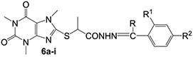
ID, structures, m.p. and yields of newly synthesized hydrazones.
|
|||||
| Compound | R | R1 | R2 | M.p. (°C) | Yield (%) |
| 6a | CH3 | H | H | 242–243 | 51 |
| 6b | CH3 | H | CH3 | 248–250 | 69 |
| 6c | CH3 | H | OCH3 | 24–249 | 73 |
| 6d | CH3 | OH | H | 239–243 | 68 |
| 6e | C2H5 | H | OH | 228–230 | 80 |
| 6f | C2H5 | H | H | 231–233 | 95 |
| 6g | C3H7 | H | H | 242–246 | 96 |
| 6h | C6H5 | H | OCH3 | 219–221 | 62 |
| 6i | CH(CH3)2 | H | H | 243–245 | 82 |
In the performed spectral analysis a slight discrepancy in the expected and the measured IR spectra for compound 6d was observed. This may be due to the intramolecular hydrogen bond which may be found in 6d resulting in formation of a stable 6-membered ring as shown in structure below (Figure

Thus no absorption bands due to the free O-H stretching vibration were found in the infrared spectra of any of the salicylidene anilines. Instead, a broad, weak band having some fine structure is found in the region from 2700 to 3100 cm-1 as is the case with 6d.
Also, a slight absorption band at 1157–1159 cm-1, which is not observed in the spectra of starting xanthines 1 and 3, is clearly visible in the spectra of studied compounds 6a-i. We consider this band to be due to N-N stretching vibration from the hydrazide residue. The identifications of this stretching vibration is a difficult task, but there is literary evidence to support our assumption (
Prediction of toxicity and drug-likeness
The toxicity and drug-likeness of compounds were predict by using PROTOX II (“ProTox-II”) web server. The ProTox-II web server provides several advantages over existing computational models. It includes both chemical and molecular target knowledge. The prediction scheme is classified into different levels of toxicity such as oral toxicity as well as organ toxicity (hepatotoxicity). ProTox-II incorporates molecular similarity and pharmacophore based fragment propensities (
An MDDRlike rule published by Oprea et al. (
The values of the calculated molecular descriptors of tested compounds 6a–i.
| Comp. | Molweight | Num. of hydrogen bond acceptors | Num. of hydrogen bond donors | Num. of atoms | Num. of bonds | Num. of rings | Num. of rotable bonds | Total charge | Molecular Polar Surface Area |
|---|---|---|---|---|---|---|---|---|---|
| Cff | 194.19 | 3 | 0 | 14 | 15 | 2 | 0 | 0 | 61.82 |
| 3 | 294.37 | 3 | 0 | 20 | 21 | 2 | 3 | 0 | 90.28 |
| 4 | 312.34 | 5 | 0 | 21 | 22 | 2 | 4 | 0 | 113.42 |
| 5 | 312.35 | 6 | 0 | 21 | 22 | 2 | 4 | 0 | 142.24 |
| 6a | 414.48 | 6 | 0 | 29 | 31 | 3 | 6 | 0 | 128.58 |
| 6b | 428.51 | 6 | 0 | 30 | 32 | 3 | 6 | 0 | 128.58 |
| 6c | 444.51 | 7 | 0 | 31 | 33 | 3 | 7 | 0 | 137.81 |
| 6d | 430.48 | 7 | 0 | 30 | 32 | 3 | 6 | 0 | 148.81 |
| 6e | 428.51 | 6 | 0 | 30 | 32 | 3 | 7 | 0 | 128.58 |
| 6f | 444.51 | 7 | 0 | 31 | 33 | 3 | 7 | 0 | 148.81 |
| 6g | 442.53 | 6 | 0 | 31 | 33 | 3 | 8 | 0 | 128.58 |
| 6h | 506.58 | 7 | 0 | 36 | 39 | 4 | 8 | 0 | 137.81 |
| 6i | 442.53 | 6 | 0 | 31 | 33 | 3 | 7 | 0 | 128.58 |
Drug-induced hepatotoxicity is a significant cause of acute liver failure and one of the major reasons for the withdrawal of drugs from the market. Drug-induced liver injury (DILI) is either a chronic process or a rare event. However, prediction of DILI is important and one of the safety concerns for the drug developers, regulators and clinicians (
The results of prediction are shown in Table
Results of toxicity prediction with ProTox-II webserver.
| Comp. | Hepatotoxicity prediction | Probability | Predicted LD50 [mg/kg b.w.] | Predicted toxicity class |
|---|---|---|---|---|
| Cff | Inactive | 0.97 | 127 | 3 |
| 3 | Active | 0.54 | 910 | 4 |
| 4 | Inactive | 0.65 | 196 | 3 |
| 5 | Active | 0.50 | 550 | 3 |
| 6a | Inactive | 0.56 | 550 | 4 |
| 6b | Inactive | 0.56 | 550 | 4 |
| 6c | Inactive | 0.53 | 150 | 3 |
| 6d | Inactive | 0.52 | 150 | 3 |
| 6e | Inactive | 0.55 | 660 | 4 |
| 6f | Inactive | 0.51 | 150 | 3 |
| 6g | Inactive | 0.54 | 790 | 4 |
| 6h | Inactive | 0.53 | 3000 | 5 |
| 6i | Inactive | 0.56 | 660 | 4 |
Prediction of substrate/metabolite specificity
The evaluated set of compounds was subjected to a prediction of substrate/metabolite specificity, using an SMP web-service (
“Probability to be Active” (Pa) values for the substrate based prediction result of 6a–i, 3, 4, 5 and Caffeine.
| Cmpnd | 1A2 | 2A6 | 2E1 | 2C8 |
|---|---|---|---|---|
| Cff | 0.912 | 0.942 | 0.921 | 0.770 |
| 3 | 0.631 | 0.654 | 0.693 | 0.772 |
| 4 | 0.650 | 0.714 | 0.701 | 0.781 |
| 5 | 0.526 | |||
| 6a | 0.672 | 0.697 | 0.687 | 0.594 |
| 6b | 0.635 | 0.732 | 0.729 | 0.606 |
| 6c | 0.679 | 0.705 | 0.695 | 0.629 |
| 6d | 0.667 | 0.521 | 0.539 | |
| 6e | 0.754 | 0.794 | 0.788 | 0.673 |
| 6f | 0.798 | 0.621 | 0.688 | 0.546 |
| 6g | 0.822 | 0.858 | 0.839 | 0.623 |
| 6h | 0.636 | 0.657 | 0.659 | 0.704 |
| 6i | 0.721 | 0.764 | 0.765 | 0.712 |
“Probability to be Active” (Pa) values for the metabolite based prediction result of 6a–i, 3, 4, 5 and Caffeine.
| Cmpnd | 1A2 | 2E1 | 3A4 |
|---|---|---|---|
| Cff | 0.944 | 0.858 | |
| 3 | 0.677 | 0.677 | |
| 4 | 0.833 | 0.504 | |
| 5 | 0.735 | 0.520 | |
| 6a | 0.808 | 0.568 | 0.554 |
| 6b | 0.818 | 0.557 | 0.544 |
| 6c | 0.855 | 0.539 | |
| 6d | 0.667 | 0.583 | |
| 6e | 0.859 | 0.686 | |
| 6f | 0.781 | 0.742 | |
| 6g | 0.903 | 0.762 | 0.515 |
| 6h | 0.833 | ||
| 6i | 0.841 | 0.637 |
In vitro evaluation of effect on isolated rat liver microsomes
It is a well known fact that Caffeine is metabolized by CYP1A2. It is of interest to establish whether during their metabolism the the newly synthesized caffeine derivatives 6a-i would be biotransformed into reactive metabolites and by this to reveal possible pro-oxidative properties.
Thus we decided to evaluate their effect on isolated rat liver microsomes, as an in vitro system, which contains most of the isoforms of the cytochrome P450 system.
Administered alone, all of the tested compounds didn’t reveal statistically significant pro-oxidant effects on isolated rat liver microsomes (Figure

4. Conclusion
The present study describes the synthesis of series new caffeine-8-(2-thio)-propanoic hydrazid-hydrazone derivatives which are subjected to in silico prediction of substrate/metabolite specificity. Based on the obtained data we consider the tested compounds to perform most probably as CYP1A2 substrates. The newly synthesized hydrazid-hydrazones were also subjected to in silico prediction of the Drug Induced Liver Injury (DILI). The prediction for all compounds indicates that they would not show hepatotoxicity. The carried out in vitro studies confirmed that the tested compounds didn’t reveal statistically significant hepatotoxicity, due to pro-oxidant effects, on sub-cellular (isolated rat liver microsomes) level. These results underlined the tested molecules as promising structures for design of future compounds with low hepatotoxicity.
References
- Acheson KJ, Gremaud G, Meirim I, Montigon F, Krebs Y, Fay LB, Gay L-J, Schneiter P, Schindler C, Tappy L (2004) Metabolic effects of caffeine in humans: lipid oxidation or futile cycling? American Journal of Clinical Nutrition 79. https://doi.org/10.1093/ajcn/79.1.40
- Al Deeb SA, AlMoutaery KA, Arshaduddin M, Biary N, Tariq M (2002) Effect of acute caffeine on severity of harmaline induced tremor in rats. Neuroscience Letter 325: 219–218. https://doi.org/10.1016/S0304-3940(02)00042-3
- Alsabri S, Mari W, Younes S, Alsadawi M, Oroszi T (2018) Kinetic and dynamic description of caffeine. Journal of Caffeine and Adenosine Research 8: 3–9. https://doi.org/10.1089/caff.2017.0011
- Banerjee B, Dehnbostel FO, Preisner R (2018a) Prediction is a balancing act: importance of sampling methods to balance sensitivity and specificity of predictive models based on imbalanced chemical data sets. Frontier in Chemistry 6: 1–11. https://doi.org/10.3389/fchem.2018.00362
- Banerjee P, Eckert AO, Schrey AK, Preissner R (2018b) ProTox-II: a webserver for the prediction of toxicity of chemicals. Nucleic Acids Research 46: W257–W263. https://doi.org/10.1093/nar/gky318
- Barcelos R, Souza M, Amaral G, Stefanello S, Bresciani G, Fighera M, Soares F, Barbosa N (2014) Caffeine supplementation modulates oxidative stress markers in the liver of trained rats. Life Sciences 96: 40–45. https://doi.org/10.1016/j.lfs.2013.12.002
- Berthou F, Guillois B, Riche C, Dreano Y, Jacqz-Aigrain E, Beaune PH (1992) variations in caffeine metabolism related to cytochrome P4501A enzymes. Xenobiotica 22: 671–680. https://doi.org/10.3109/00498259209053129
- Bezhentsev VM, Tarasova OA, Dmitriev AV, Rudik AV, Lagunin AA, Filimonov DA, Poroikov VV (2016) Computer-aided prediction of xenobiotics metabolism in the human organism. Russ Chem Rev 85: 854–879. https://doi.org/10.1070/RCR4614
- Bharanidharan S, Saleem H, Subashchandrabose S, Suresh M, Ramesh Babu N (2017) FT-IR, FT-Raman and UV-Visible Spectral Analysis on (E)-N’-(thiophen-2-ylmethylene) Nicotinohydrazide. Archives in Chemical Research 1: 1–14. https://doi.org/10.21767/2572-4657.100007
- Bloomer JC, Clarke SE, Chenery RJ (1995) Determination of P4501A2 activity in human liver microsomes using [3-’4C-methyl]caffeine. Xenobiotica 25: 917–927. https://doi.org/10.3109/00498259509046663
- Chen M, Suzuki A, Thakkar S, Yu K, Hu C, Tong W (2016) DILIrank: The largest reference drug list ranked by the risk for developing drug-induced liver injury in humans. Drug Discovery Today 21: 648–653. https://doi.org/10.1016/j.drudis.2016.02.015
- Deby C, Goutier R (1990) New perspectives on the biochemistry of superoxide anion and the efficiency of SOD. Biochem Pharmacol 39: 399–405. https://doi.org/10.1016/0006-2952(90)90043-K
- Devasagayam TP, Kamat JP, Mohan H, Kesavan PC (1996) Caffeine as an antioxidant: inhibition of lipid peroxidation induced by reactive oxygen species. Biochimica et biophysica acta 1282: 63–70. https://doi.org/10.1016/0005-2736(96)00040-5
- Dianzani MU, Muzio G, Biocca ME, Canuto RA (1991) Lipid peroxidation in fatty liver induced by caffeine in rats. International Journal of Tissue reactions 13: 79–85.
- Franco R, Onatibia-Astibia A, Martinez-Pinilla E (2013) Health benefits of methylxanthines in cacao and chocolate. Nutrients 5: 4159–4173. https://doi.org/10.3390/nu5104159
- GHS-unece (2019) GHS-unece. http://www.unece.org/trans/danger/publi/ghs/ghs_welcome_e.html [accessed January 29.2019]
- Guengerich FP (1989) Analysis and characterizaton of enzymes. In: Hayes AW (Ed.) Principles and methods of toxicology. New York Raven Press, 777–814.
- Gulaczyk I, Krezglewski M, Valentin A (2003) The N–N stretching band of hydrazine. Journal of Molecular Spectroscopy 220: 132–136. https://doi.org/10.1016/S0022-2852(03)00106-1
- Gulcin I (2008) In vitro prooxidant effect of caffeine. Journal of Enzyme Inhibition and Medicinal Chemistry 23: 149–152. https://doi.org/10.1080/14756360701306404
- Hetzler RK, Knowlton RG, Somani SM, Brown DD, Perkins RM (1990) Effect of paraxanthine on FFA mobilization after intravenous caffeine administration in humans. Journal of Applied Physiology 68: 44–47. https://doi.org/10.1152/jappl.1990.68.1.44
- Hristova-Avakumova N, Yoncheva K, Nikolova-Mladenova B, Traykov T, Momekov G, Hadjimitova V (2017) 3-Methoxy aroylhydrazones – free radicals scavenging, anticancer and cytoprotective potency. Redox Report 22: 408–417. https://doi.org/10.1080/13510002.2016.1276256
- Kolayli S, Ocak M, Kucuk M, Abbasoglu R (2004) Does caff eine bind to metal ions? Food Chemistry 84: 383–388. https://doi.org/10.1016/S0308-8146(03)00244-9
- Kot M, Daniel WA (2008a) The relative contribution of human cytochrome P450 isoforms to the four caffeine oxidation pathways: an in vitro comparative study with cDNA-expressed P450s including CYP2C isoforms. Biochemical Pharmacolology 76: 543–551. https://doi.org/10.1016/j.bcp.2008.05.025
- Kot M, Daniel WA (2008b) Relative contribution of rat cytochrome P450 isoforms to the metabolism of caffeine: the pathway and concentration dependence. Biochemical Pharmacolology 75: 1538–1549. https://doi.org/10.1016/j.bcp.2007.12.017
- Liu J, Mansouri K, Judson RS, Martin MT, Hong H, Chen M, Xu X, Thomas RS, Shah I (2015) Predicting hepatotoxicity using ToxCast in vitro bioactivity and chemical structure. Chemical Research in Toxicology 28: 738–751. https://doi.org/10.1021/tx500501h
- Lowry OH, Rosebrough NJ, Farr AL, Randall RJ (1951) Protein measurement with the Folin phenol reagent. Journal of Biological Chemistry 193: 265–275.
- Metro D, Cernano V, Santoro D, Papa M, Buemi M, Benvenga S, Manasseri L (2017) Beneficial effects of oral pure caffeine on oxidative stress. Journal of Clinical & Translational Endocrinology 10: 22–27. https://doi.org/10.1016/j.jcte.2017.10.001
- Mitkov J, Georgieva M, Zlatkov A (2012) Development of an optimized synthetic approach for synthesis of caffeine-8-thioglycolic acid and its ester derivatives. Pharmacia 59: 17–23.
- Mitkov J, Kasabova-Angelova A, Kondeva-Burdina M, Tzankova V, Tzankova D, Georgieva M, Zlatkov A (2019) Design, Synthesis and Evaluation of 8-Thiosubstituted 1,3,7-Trimethylxanthine Hydrazones with In Vitro Neuroprotective and MAO-B Inhibitory Activities. Medicinal Chemistry 15. https://doi.org/10.2174/1573406415666190531121927
- Nikolaevskii AN, Kniga OP, Khizhan EI, Tikhonova GA, Vinogradov VV, Khizhan AI (2012) Antioxidant activity of hydrazones with sterically hindered phenol fragments. Russian Journal of Physical Chemistry A 86: 1816–1820. https://doi.org/10.1134/S0036024412120205
- Oprea TI (2000) Property distribution of drug-related chemical databases. Journal of Computer-Aided Molecular Design 18: 251–264. https://doi.org/10.1023/A:1008130001697
- Oprea TI, Gottfries J, Sherbukhin V, Svensson P, Kiihler TC (2000) Chemical information management in drug discovery: Optimizing the computational and combinatorial chemistry interfaces. Journal of Molecular Graphics and Modelling 18: 512–524. https://doi.org/10.1016/S1093-3263(00)00066-8
- Pasaoglu H, Demir FEO, Demirtas CY, Hussein A, Pasaoglu OT (2011) Th e eff ect of caff eine on oxidative stress in liver and heart tissues of rats. Turkish Journal Of Medical Sciences 41: 665–671.
- ProTox-II (2019) ProTox-II. http://tox.charite.de/protox_II [accessed January 28.2019]
- Sarıgöl D, Yüksel D, Okay G, Uzgören-Baran A (2015) Synthesis and structural studies of acyl hydrazone derivatives having tetrahydrocarbazole moiety. Journal of Molecular Structure 1086: 146–152. https://doi.org/10.1016/j.molstruc.2014.12.092
- Shi X, Dalal NS, Jain AC (1991) Antoxidant behaviour of caffeine: efficient scavenging of hydroxyl radicals. Food and Chemical Toxicology 29: 1–6. https://doi.org/10.1016/0278-6915(91)90056-D
- Thakkar S, Chen M, Fang H, Liu Z, Roberts R, Tong W (2018) The Liver Toxicity Knowledge Base (LKTB) and drug-induced liver injury (DILI) classification for assessment of human liver injury. Expert Review of Gastroenterology and Hepatology 12: 31–38. https://doi.org/10.1080/17474124.2018.1383154
- Welsh E, Bara A, Barley E, Cates CJ (2012) Caffeine for asthma. John Wiley & Sons, Ltd. https://doi.org/10.1002/14651858.CD001112.pub2