|
Corresponding author: Taras Chaban ( chabantaras@ukr.net ) Academic editor: Georgi Momekov
© 2020 Olha Ripetska, Anatoliy Mahlovanyy, Ihor Hrynovets, Taras Chaban, Volodymyr Hrynovets, Anna Buchkovska, Stefan Harkov, Ihor Deneha, Oksana Pasko, Marta Renka.
This is an open access article distributed under the terms of the Creative Commons Attribution License (CC BY 4.0), which permits unrestricted use, distribution, and reproduction in any medium, provided the original author and source are credited.
Citation:
Ripetska O, Mahlovanyy A, Hrynovets I, Chaban T, Hrynovets V, Buchkovska A, Harkov S, Deneha I, Pasko O, Renka M (2020) Clinical study of the efficacy of si-containing polishing paste for the professional hygiene procedures in patients with periodontal disease. Pharmacia 67(1): 17-22. https://doi.org/10.3897/pharmacia.67.e35152
|
Abstract
Silicon-containing (Si-containing) polishing paste has been tested clinically in patients with chronic generalized periodontitis, I-st degree of severity. All patients have been examined for the presence of supra- and sub-gingival calculus and bleeding on probing on a six- month-testing time and within one year after conducting the professional hygienic procedures. It has been revealed that the proposed Si-containing polishing paste proved to be effective in the maintenance of gingival health in the lapse of time from 1 to 12 months after treatment. The majority of patients showed neither dental deposits nor bleeding on probing up in the interval from 1 to 12 months after dental scaling and polishing with Si-containing polishing paste.
Keywords
bleeding, dental calculus, dental plaque, dental scaling, generalized periodontitis, Si-containing polishing paste
Introduction
Providing medical care to a dental patient with a diagnosis of inflammatory periodontal disease, regardless of etiological factors, involves an instrumental procedure - the removal of dental calculus – scaling, as well as leveling and polishing the surface – root planning of the “cervix dentis” and “radix dentis” (
The clinical evaluation of the feasibility and effectiveness of polishing with Si-containing polishing paste, which was used at the final stage of the dental scaling procedure, after the treatment of the root surface of tooth, and with the additional use of standard Gray curettes, in combination which the Si-containing polishing paste was performed in our investigations (
Experimental part
Materials and methods
93 patients with chronic generalized periodontitis, I-st stage of severity (the depth of periodontal pockets up to 3,5 mm) were under study. All periodontal patients were previously examined for the presence of dental plaque and calculus under the gums and gingival bleeding during the probing. All patients were provided with professional hygiene procedure of root scaling, planning and polishing.
Patients were divided into two groups (the main and the control): The main group numbered 32 patients (16–29 years old) and 24 patients (30–55 years old). The control group included: 17 patients (16–29 years old) and 20 patients (30–55 years old). The procedure of removing the dental deposits from the root surfaces of the teeth (scaling) was performed for patients of both groups.
In patients from the main group, the cervical zone of the teeth was treated, there to, with Si-containing polishing paste.
Subsequently, for all patients, a basic treatment course was performed in accordance with the requirements of the Order of the Ministry of Health Care of Ukraine No 566 “On Approval of Medical Assistance Protocols” (
Results and discussion
The results of the study include indicators of clinical evaluation – the feasibility and effectiveness of the use of Si-containing polishing paste, which was additionally used to polish the root on the necks of teeth in patients with periodontitis of the I-st degree of severity (
Before the study, all patients were given a sanitary examination of the oral cavity with an analysis of the affected parts of the periodontal tissues, the presence of dental plaque in the form of a firm plaque under the gums and gingival bleeding during the probing.
Subsequently, for all patients, a principal course of treatment was performed in accordance with the requirements of the Order of the Ministry of Health of Ukraine No 566 “On Approval of Medical Assistance Protocols”(The Order of the Ministry of Health of Ukraine № 566 dated November 23, 2004).
In order to carry out a study aiming to improve the course of treatment, 56 patients were diagnosed with periodontitis, the I-st degree of severity (the depth of periodontal pockets up to 3,5 mm).
In view of age the patients were divided into two groups the main and the control. The main group included 32 patients (16–29 years old) and 24 patients (30–55 years old).The control group (groups for comparison) numbered: 17 patients (16–29 years old old) and 20 patients (30–55 years old old).
The procedure for removing the deposits from the root surface of the tooth was performed for patients of both groups using the standard Gray curettes. For patients from the main group, the cervical zone of the teeth was treated there to with Si-containing polishing paste. The results are shown in the Table
Dependence of the number of periodontal pockets in patients with a diagnosis – generalized periodontitis (I-st degree of gravity).
| Groups of patients | Patients with generalized periodontitis by age / years old | Number of patients | Number of periodontal pockets | |||
|---|---|---|---|---|---|---|
| Detected during examination of patients | Surveyed areas of periodontal pockets with control | |||||
| 1 month | 6 months | 12 months | ||||
| Main (Si-containing polishing paste usage) | 16–29 | 32 | 599 | 596 | 593 | 585 |
| 30–55 | 24 | 541 | 535 | 529 | 524 | |
| Control | 16–29 | 17 | 296 | 293 | 287 | 277 |
| 30–55 | 20 | 455 | 446 | 436 | 427 | |
Control over the results obtained in the lapse of time after the procedure of dental scaling and polishing of the teeth:
After 1 month, in patients aged 16–29 years old, from the main group (Table
The results of the studies confirm that in the main group of patients, if compared with the control group, the criteria for the treatment provided are significantly better (75 % to 48,3 %). The analysis of the obtained data results of a 6 month rehabilitation has reveal the following: in the main group of patients, 72,5%(± 1,8) cases with no bleeding, in the control group – 53,3% (± 2,9). The difference between the two groups was significant at this time (t = 5,6; p <0,01), which gives grounds to argue that the use of Si-containing polishing paste makes the gingival and roots condition of affected teeth within 6 months generally better.
After 12 months, the number of sites with periodontal absence of bleeding on probing dropped both in the main and control groups (22% (± 1,9) and 20% (± 2,3). In accordance with obtained data, there was practically no difference between the indices in patients of the main and control groups (t = 0,7; p <0,1). This indicates that after 12 months the impact of scaling and polishing on marginal periodontal condition is not manifested. The results are presented in the Table
The severity of bleeding when sensing in patients, aged 16–29 years old, with generalized periodontitis (I-st degree of gravity).
| Number of periodontal pockets in patients | Observation of bleeding gums through time periods after: | ||||||
|---|---|---|---|---|---|---|---|
| Groups ofpatients | Magnitude assessments | 1 month | 6 months | 12months | |||
| no bleeding | Bleeding on probing | no bleeding | Bleeding on probing | no bleeding | Bleeding on probing | ||
| Main si-containing polishing paste usage) | absolute | 447 | 149 | 430 | 163 | 129 | 456 |
| in, % | 75.0 ± 1.8 | 25.0 ± 1.8 | 72.5 ± 1.8 | 27.5 ± 1.8 | 22.0 ± 1.7 | 78.0 ± 1.7 | |
| Control | absolute | 142 | 151 | 153 | 134 | 55 | 222 |
| in, % | 48.3 ± 2.9 | 51.7 ± 2.9 | 53,3±2,9 | 46.7 ± 2.9 | 20.0 ± 2.4 | 80.0 ± 2.4 | |
| t | 7.8 | 5.6 | 0.7 | ||||
| p | <0.01 | <0.01 | >0.1 | ||||
In patients with acute form of generalized periodontitis, I-st degree of severity, 30–55 years of age (Table
After 6 months, the positive impact of the procedure of dental scaling and polishing was obvious, saving significant difference observed in the obtained results. Areas of bleeding on probing in the main and control groups were 67,2% (± 2,0) and 34,4% (± 2,2) respectively. The polishing of the exposed tooth neck significantly prevents both, gum bleeding and exacerbation of inflammation for 6 months. The results are shown in Table
Expression of bleeding when sensing in patients, aged 30–55 years old, with generalized periodontitis (I-st degree of gravity).
| Number of periodontal pockets in patients | Observation of bleeding gums through time periods after: | ||||||
|---|---|---|---|---|---|---|---|
| Groups ofpatients | Magnitude assessments | 1 month | 6 months | 12months | |||
| no bleeding | Bleeding on probing | no bleeding | Bleeding on probing | no bleeding | Bleeding on probing | ||
| Main (with usage a si-containing polishing paste) | absolute | 505 | 30 | 355 | 174 | 90 | 434 |
| in, % | 94.4 ± 1.0 | 5.6 ± 1.0 | 67.2 ± 2.0 | 32.8 ± 2.0 | 17.2 ± 1.6 | 82.8 ± 1.6 | |
| Control | absolute | 238 | 208 | 150 | 286 | 95 | 332 |
| in, % | 53.3 ± 2.4 | 46.7 ± 2.4 | 34.4 ± 2.3 | 65.6 ± 2.3 | 22.2 ± 2.0 | 77.8 ± 2.0 | |
| p | <0.01 | <0.01 | <0.01 | ||||
The younger patients (16–29 years old) in the main group, with periodontitis, I-st degree of severity and in the older group (30–55 years old), experienced low rates of gum bleeding on probing of periodontal pockets for 6 months, if compared with control group, showed a positive effect of polishing with Si-containing polishing paste on the gums. Within 12 months after dental scaling and polishing procedures, the percentage of periodontal pockets with severe bleeding in both age groups increases, but bleeding in the younger age category was more pronounced. This tendency was maintained in a month and in 6 months. Obviously, this happens due to the predominantly acute course of the process in patients 16–29 years of age.
The effect of polishing was also studied on the exposed root cement and formation of sub-gingival calculus in patients with periodontitis, Ist stage of severity aged 16–29 years old and 30–35 years old. The obtained data are shown in the Tables
The results of the study of the preventive effect of polishing on the exposed root cement of teeth on the formation of dental calculus, in patients with periodontitis, I-st stage of severity, 30–55 years old are shown n in Table
One month after calculus removal and subsequent polishing of the exposed root cement in the main and control groups of patients, the absence of dental calculus was 91,7% (± 1,2) and 82,2% (± 1,8) respectively, and this difference was significant (t = 4,3; p ± 0,01).
After 6 months, the calculus free zones in the main group of patients reached 85,0% (± 1,5), and in the control group – 67,8% (± 2,2), which is also characterized by a significant difference (t = 4,3; p ± 0,01). On the basis of the obtained results, we can conclude as to the efficacy of scaling and polishing with Si-containing polishing paste in preventing the formation of a dental calculus for at least 6 months.
After 12 months, the percent age of sites with dental calculi in the main group of patients was 71,7% (± 1,9), and in the control group – 66,7% (± 2,2), The difference between the two indices was insignificant (t = 1,7; p ± 0,05).
Comparison of the results regarding recurrent periodontal disease in periodontitis, I-st degree of severity in patients of two age groups (Tables
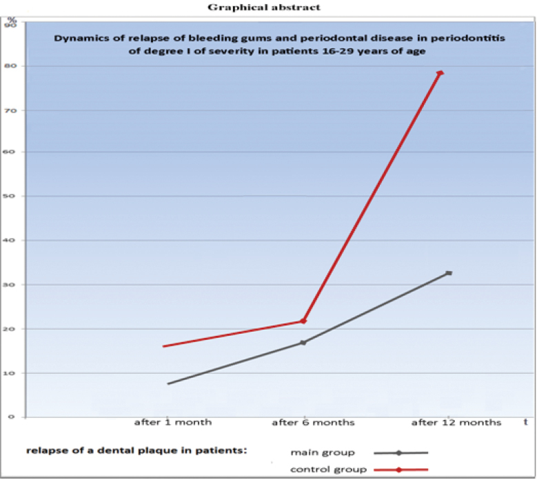
Further comparison of the data of the simultaneous presence of gingival bleeding and dental calculus after scaling with the subsequent polishing of the root surface in patients of both age categories with periodontitis I degree of severity showed that in a month period the percentage of calculus free sites and no bleeding is lower than percentage of calculus free sites. Such a pattern can be traced in all patients, but in the main group, these indicators are much better. For example, in the age group of 16–29 years, there were 94,2% of sites without dental calculi and 75% of sites with bleeding. In the control group, 83% and 48,3% of the sites, respectively. After 6 months, the majority of patients in the main group were deprived of calculus 85,8%, and bleeding was observed in 72,5% of the sites, while in the control group -75% and 53,3% of the sites, respectively. After 12 months in both groups, with a fairly significant percentage of calculus-free sites, there is a low percentage of sites without bleeding. The results are presented in the Fig.
Frequency of detection of a dental plaque under the gums of the teeth during periodontitis (I-st degree of gravity) in patients aged 16–29 years old.
| Number of periodontal pockets in patients | Observation of bleeding gums through time periods after: | ||||||
|---|---|---|---|---|---|---|---|
| Groups ofpatients | Magnitude assessments | 1 month | 6 months | 12months | |||
| no bleeding | Bleeding on probing | no bleeding | Bleeding on probing | no bleeding | Bleeding on probing | ||
| Main (with us age a si-containing polishing paste) | absolute | 561 | 35 | 509 | 84 | 356 | 229 |
| in, % | 94.2 ± 0.3 | 0.8 ± 0.3 | 85.8 ± 1.4 | 14.2 ± 1.4 | 60.8 ± 2.0 | 39.2 ± 2.0 | |
| Control | absolute | 243 | 50 | 215 | 72 | 157 | 120 |
| in, % | 83.0 ± 2.2 | 17.0 ± 2.2 | 75.0 ± 2.5 | 25.0 ± 2.5 | 56.7 ± 3.0 | 43.3 ± 3.0 | |
| t | 5.1 | 3.7 | 1.17 | ||||
| p | <0.01 | <0.01 | >0.1 | ||||
Frequency of detection of a dental plaque under the gums of the teeth during periodontitis (I-st degree of gravity) in patients aged 30–55 years old.
| Number of periodontal pockets in patients | Observation of bleeding gums through time periods after: | ||||||
|---|---|---|---|---|---|---|---|
| Groups ofpatients | Magnitude assessments | 1 month | 6 months | 12months | |||
| no bleeding | Bleeding on probing | no bleeding | Bleeding on probing | no bleeding | Bleeding on probing | ||
| Main (with usage a si-containing polishing paste) | absolute | 491 | 44 | 450 | 79 | 376 | 148 |
| in, % | 91.7 ± 1.2 | 8.3 ± 1.2 | 85.0 ± 1.5 | 15.0 ± 1.5 | 71.7 ± 1.9 | 28.3 ± 1.9 | |
| Control | absolute | 367 | 79 | 296 | 140 | 285 | 142 |
| in, % | 82.2 ± 1.8 | 17.8 ± 1.8 | 67.8 ± 2.2 | 32.2 ± 2.2 | 66.7 ± 2.2 | 33.3 ± 2.2 | |
| t | 4.3 | 6.4 | 1.17 | ||||
| p | <0.01 | <0.01 | >0.1 | ||||

Dynamics of gingival bleeding in patients 30–55 years old with periodontitis (I-stage of heaviness) in the main and control groups. *On the horizontal axis – Repeated r periods of time: 1) -1 month; 2)-6 months; and 3) -12 months. *On the vertical axis: Quantity of periodontal areas with bleeding on probing (%).

Dynamics of recurrence of subclinical calculus in patients 16–29 years with periodontitis (I stage of heaviness). *On the horizontalaxis – Repeated examinations in periods of time: 1) - 1 month; 2) - 6 months; and 3) - 12 months. *On the vertical axis: Quantity of periodontal areas with bleeding on probing (%).

Dynamics of recurrence of gum bleeding and periodontal inflammation in patients 16–29 years with periodontitis (I stage of heaviness). *On the horizontal axis – Repeated examination in periods of time: 1) -1 month; 2)-6 months; and 3) -12 months. *On the vertical axis: Quantity of periodontal areas with bleeding on probing (%).
On the basis of comparative analysis of the data of bleeding and the apparent calculus in the examined patients both, of control and main groups, one can conclude of positive effect due to applied abrasive and antiseptic paste.
Accordingly, having analyzed the indicators of bleeding and the presence of sub-gingival calculus in patients with mild periodontitis starting from the age of 16 to 29 years, we can draw a conclusion that the number of sites with no bleeding is not the same as the number of calculus- free sites. In all analyzed cases, the percentage of sites without calculus was higher than the percentage of sites with no bleeding. This pattern is traced in a month, six months and a year periods after the procedure of dental scaling and polishing, in both, the main and control groups.
Conclusions
- The obtained data allow to state that the condition of gums depends not only on the presence of the sub-gingival deposits, since there is a certain percentage of dental-free areas with signs of bleeding. In the main group, however, in comparison with the control group, there were significantly higher indicators, both in the number of areas without bleeding, and in calculus-free areas.
- The results also confirm the effectiveness of polishing of the exposed root cement for improving the outcome of treatment in patients with I stage of periodontitis. However, it should be noted that the processing of cement even with polishing is not always effective as to the elimination of bleeding symptoms. Obviously, polishing is more effective in preventing the formation of new dental deposits.
- From the analysis of the obtained data, it can be concluded that polishing has a positive effect on gingival condition, and this effect is noticeable within 6 months after polishing. However, polishing has more continuos effect on the prevention of dental deposits than on preventing gingival bleeding.
References
- AlJehani YA (2014) Risk factors of Periodontal disease: Review of the Literature. International Journal of Dentistry 2: 1–9. https://doi.org/10.1155/2014/182513
- Drisko CA (1998) Root toolkit Power-driven versus manuals calers, which one. Dental Clinics of North America 42(2): 229–244.
- Garranza FA, Newman MG (2016) Clinical periodontology. 10-thed. W.B. Saunders Co. 2016, 458.
- Grokholsky AP, Kodol NA, Tsentilo TD (2000) Nazudeposits: their in fluence on teeth, perineum tissues and organism. Kyiv, 160.
- Heorgiev VI (2002) Professional oral hygiene in periodontology. Dentist 2002(2): 47–49.
- Herbert F, Ulrich P (2007) Sacksper Periodontitis. GalDent, 40.
- Hrynovets I, Mahlovanyy A, Deneha I, Ripetska O, Hrynovets V, Buchkovska A (2016) Аpplication of Different Medicinal Forms in Dental Practice. Danylo Halytsky National Medical Universit, 105.
- Mahlovanyy A, Hrynovets I, Ripetska O, Hrynovets V, Deneha I (2015) Medications Commonly Usedin Conservative Dentistry. Education-methodological manual. Lviv, 104.
- Müller HP (2004) Periodontology. Lviv: GalDent, 158–167.
- Ripetska OR, Kuhta VS (2000) Estimation of the surface of the teeth cementin the process of eliminating dental deposits and polishing. News of dentistry 2(23): 59–60.
- Ripetska O, Deneha I, Hrynovets V, Hysyk M (2004) Diseases of the Periodontium. Etiology. Pathogenesis Diagnosis Treatment. Lviv-Press, 174.
- Sandhu HS, Salloum IA, Stakiw JE (1998) Scaling and Root Planing: Handversus Power-driven Instruments. General Dental 64(4): 269–275.
- Ministry of Health of Ukraine (2004) The Order of the Ministry of Health of Ukraine № 566 dated November 23, 2004 “Onapproval of medical treatment protocols in the specialties”, orthopedic stomatology”, “therapeutic stomatology”, “surgical stomatology”, “orthodontics”, “pediatric therapeutic stomatology”, “pediatric surgical stomatology”. ACS Publications Division Home Page. http://old.moz.gov.ua/ua/portal/dn_20041123_566.html [Accessed November 2004]