|
Corresponding author: Maryan Lelyukh ( lelyukh.m@gmail.com ) Academic editor: Georgi Momekov
© 2020 Viktoria Dovhanyk, Anatoliy Mahlovanyy, Stefan Harkov, Volodymyr Synytsa, Volodymyr Hrynovets, Ihor Hrynovets, Ihor Chaban, Maryan Lelyukh.
This is an open access article distributed under the terms of the Creative Commons Attribution License (CC BY 4.0), which permits unrestricted use, distribution, and reproduction in any medium, provided the original author and source are credited.
Citation:
Dovhanyk V, Mahlovanyy A, Harkov S, Synytsa V, Hrynovets V, Hrynovets I, Chaban I, Lelyukh M (2020) Spectroscopic criteria for early diagnosis of changes in the mineral and organic matrix of hard dental tissues. Pharmacia 67(1): 5-12. https://doi.org/10.3897/pharmacia.67.e35126
|
Abstract
At the present stage of development of medical science, experimental and clinical dentistry is often used in scientific research, which allows diagnosing preclinical stages of manifestations of changes in tissues and organs of the oral cavity and biological fluids at the level of nanotechnology enabling to study the mechanisms of molecular transformation that lead to occurrence of pathological changes. One of the promising methods of research is infrared spectroscopy. The advantage of the method is that the absorption spectrum can be obtained with a small amount of the test substance (0.1–0.01 cm3) and under conditions of different aggregate states (solution, powder, liquid).
Graphical abstract
Keywords
infrared spectroscopy, hydroxyapatite, carbamide peroxide
Introduction
Infrared spectroscopy (IRS) is a part of molecular optical spectroscopy that studies the absorption and reflection spectra of electromagnetic radiation in the infrared region. This method can be used to study the state of the mineral and organic matrix of hard dental tissues and the effect of factors of exogenous and endogenous origin, as well as the state of indicators of oral fluid that reflect changes in the structure of teeth, periodontal and oral tissues, and for the purpose of planning the volume of dental intervention with further evaluation of the treatment efficacy.
The molecular structure of apatites – mineralized tissues of the tooth, can differ sharply both in the conditions of the physiological norm and in pathological conditions. The optimal composition of the hydroxyapatite corresponds to the formula Ca10 (PO4)6 (OH)2 with a molecular ratio of Ca/P ions corresponding to 1.67 (
It is established that the nature of chemical bonds in the enamel structure allows determining the initial changes that lead to a violation of the stoichiometry of hydroxyapatite crystals, as well as its demineralization and recrystallization (
There are two types of oscillations of the molecules: valence (oscillations of atoms occur along the axis of the valence bond) and deformation (the atoms in the oscillations deviate from the axis of the valence bond). For each chemical group, there are the corresponding oscillation frequencies (wave numbers) and absorption bands (characteristics). On the spectra, fluctuations of structural molecular and ionic units of the inorganic matrix such as phosphate, carbonate, and hydrophosphate ions are recorded, as well as numerous oscillations associated with a protein matrix. The spectrum band is described by the following indicators: intensity, width, type of polarization (
Experimental part
Materials and methods
The main aim of the work was to investigate the enamel discoloration of vital teeth and to determine the effect of remineralizing therapy on valence grouping of the enamel surface of the experimental teeth with the help of the active ingredient of carbamide peroxide (СP), as well as to apply the IRS in order to confirm the effectiveness of the therapeutic correction results.
Results and discussion
As a result of analysis of scientific sources, short reports, patents and own research (
carbamide peroxide – 30
glycerol – 37
vinylin – 18
aerosol – 10
quercetin – 4
three sodium ethylene
diamine tetra acetate – 1
For dental bleaching at home, the composition is:
carbamide peroxide – 15
glycerol – 46
vinylin – 22
aerosol – 12
quercetin – 4
three sodium ethylene
diamine tetra acetate – 1
The components of the two prescriptions are compatible with each other, and their quantitative and qualitative characteristics correspond to the concentrations described in the Pharmacopoeial Articles, the Temporary Pharmacopoeial Articles, the specifications and the instructions for use.
Unlike most of the bleaching agents, the above-mentioned agents have a pH of 6.8–7.1, which prevents the demineralization of the surface layer of enamel. The bleach was kept for 20 days in a light-proof package.
The СP solution is unstable and is applied to the surface of the tooth and decomposes into hydrogen peroxide (up to 30% by weight of СP) and carbamide (70%). In the further decay of hydrogen peroxide, water and oxygen are formed, and urea disintegrates into ammonia and СP. These molecular compounds freely penetrate the surface structures of the enamel due to the relatively small molecular weight (34 g/mol for hydrogen peroxide and 60 g/mol for urea). One of the breakdown products of СP is free hydroxide radicals that can inactivate enzymes, alter the structure of macromolecules and disrupt cells and intracellular organelles. Therefore, when developing the composition of bleaching therapeutic compositions, they should include antioxidant agents that prevent the harmful effects of high-level hydroxide radicals, protecting the functional groups of proteins and other bioactive molecules of organs and tissues of the oral cavity. For this purpose quercetin, an active pharmaceutical ingredient with a pronounced antioxidant effect is included in the proposed bleaching compositions.
It should also be noted that carbamide СО(NH2)2 neutralizes acids (primarily milk) that produce microorganisms of the dental plaque. This process is due to the cleavage of the urea with the enzyme with the formation of carbon dioxide (CO2) and ammonia (NH3). Since NH3 has an alkaline environment, it neutralizes the acid. The presence of aerosil composition, which is silicon sorbent, prolongs the action of the active component – urea peroxide and its gradual removal from the therapeutic form. Vinylin is known for its anti-inflammatory, enveloping, and regenerating effect, which also reduces the irritant effect of urea peroxide on gum in the formulation. 3-natrium EDTA was introduced into the bleaching composition as a stabilizer that prevents catalytic decomposition of urea peroxide.
At the initial stage, the vestibular surface of the enamel of intact teeth treated by the method of home (14 teeth) and clinical (12 teeth) bleaching in patients aged 17–35 years old was removed by orthodontic indexes. The oral surface of the same experimental teeth served as control.
After the last bleaching procedure, the teeth to be prepared were purified by the end of the brush and washed with distilled water. Enamel was ground with a diamond bur at a rate of 2000 rpm. Overheating of solid tissues was prevented by periodic irrigation of tissues with distilled water and by drying them in a stream of air, as well as by intermittent loading of a rotating tool on a tooth. The sample of enamel was thoroughly triturated in an agate mortar and sieved through a sieve with a diameter of holes of 0.04 mm. To remove moisture and to prevent the chemical activity of potassium bromide (KBr) against hydroxyapatite, the powder was dried at 100–105 °C for 3 hours in a drying cabinet. A hard powder of enamel (3 mg) was mixed in an agate mortar with 300 mg of potassium bromide, the particle size of which was about 30 ... 40 microns. The obtained samples were the same mass, which made it possible to quantify the relative optical density of absorption bands of the IRS, in particular, to determine the content of the test substance in the sample. The samples were prepared by compression of KBr tablets under pressure of 1200–1500 kg/cm2 under vacuum conditions. Infrared absorption spectra of enamel were obtained in the range of 4000–400 cm-1 on the Avatar 320 FT-IR IRS meter (“Nicolet", Japan) with Fourier transform, which allows more accurate determination of the nature of experimental objects (
After obtaining the results, the parameters of each individual absorption band were analyzed and the concentration of the substance in the samples was determined. The analysis was based on the dependence of the intensity of absorption bands on the concentration of the test substance according to the Beer-Lambert-Bouguer formula (
, (1)
at that D – optical density; lg – logarithm; g – the intensity of the incident beam of light; g0 – the intensity of the passage of radiation.
In order to process and analyze the results obtained, methods of variation statistics were used: the estimation of the difference between the frequency of occurrence of the characteristic in separate series of observations, the comparison of serial values and the mean square deviation. To assess the significance of the difference in the indices of the two populations obtained during the study, the degree of difference between their mean scores was determined using the Student’s t-test. The difference in statistical variables was considered significant at p<0.05. “Statistica for Windows" advanced analytics software, “Excel Statistica 7.0" spreadsheets (“Microsoft", USA) were used.
An analysis of the results of infrared spectra of intact enamel revealed the types of oscillations of valence grouping of organic and mineral components characteristic of enamel. In the range of 4000 ... 400 cm-1 (Fig.
565 cm-1 and 603 cm-1 – asymmetric deformation vibrations ν4 (PO43-)
872 cm-1 – non-expressed vibrations ν2 (СО32-)
960 cm-1 – complete metric valence fluctuations ν1 (PO43-)
1040 cm-1 and 1085 cm-1 – asymmetric valence fluctuations ν3 (PO43-)
1417 cm-1, 1457 cm-1 – asymmetric flat valence vibrations ν3 of СО32- ion
1540 cm-1 – Amide II valence oscillations (NH)
1650 cm-1 – Amide I and valence oscillations (C-O)
2849 cm-1 and 2920 cm-1 – valence fluctuations (C-H), (NH)
3430 cm-1 – oscillations of ОН-group of molecules of tightly bound water
In assessing the IRS of the enamel of the tooth, the band 1100–900 cm-1 is the most intense, which characterizes the valence fluctuations of the phosphate anion PO43-. In the complex contour of this band, three maxima are distinguished: 1040 cm-1 – the highest, 1085 cm-1 – medium, 960 cm-1 – the weakest intensity. Along with the absorption bands inherent in the anion PO43-, there are 1090–1030 cm-1 bands characteristic for the two-substituted orthophosphate НРО42-, which are weak intensity due to the low content of soluble phosphate forms. In addition, the composition of hydroxyapatite crystals compounds includes Me*CO3 (900–850 cm-1). Comparison of the contents of Me*CO3 with phosphates is insignificant. In the region of the oscillations of the connections of the anion СО32-, absorption bands are determined due to valence vibrations ν1 (1430–1405 cm-1, 1460–1450 cm-1), as well as valence vibrations ν2 (880–870 cm-1). The analysis of the strips of compounds СО32- determines the nature of the mineralization of enamel, that is, the low content of carbonates indicates high mineralization of enamel.

The valence fluctuations of the amide groups of proteins are in the range of 2920–2860 cm-1, 2390–2370 cm-1, and 1700–1500 cm-1. The valence fluctuations of NH groups are the most important among them, which are designated as Amide II and the carbonyl strip Amid I. The valence vibrations in the region of 2920–2860 cm-1 belong to weakly evident valence fluctuations of CH- and NH-groups. Valid oscillations of Amide II, with a maximum at 1540 cm-1, are due to plane fluctuations accompanied by the rotation of the N-H bond. Therefore, the band Amide I, with a maximum at 1660 cm-1 (carbonyl), is characteristic of oscillations, in which the length of the C-O bond changes. The position of these bands is a function of conformation of the protein, in addition, they are connected with deformation oscillations of the bond O = C-N-H. The absorption band of Amide II band exceeds the absorption area of Amide I, probably due to the absence of collagen proteins in the enamel. The protein matrix of the enamel is represented by the three-dimensional protein-calcium-protein structure, the basis of which is glycosaminoglycans and Ca2+ ions. The mature, formed enamel almost does not contain proline and oxyproline (
The absorption bands of groups that have hydrogen atoms are located at the short-wave end of the fundamental frequency of oscillations of 4000–2000 cm-1 molecules. The greatest frequencies are characterized by the band of valence fluctuations of free hydroxide groups, which are in the region of 3650–3590 cm-1. The absorption band with a maximum at 3340 cm-1 relates to oscillations of strongly coupled water molecules (OH-) (
Thus, the mineral enamel matrix is mainly composed of insoluble forms of trisubstituted calcium phosphate Ca3 (PO4)2, which is part of the hydroxyapatite. In addition, valence oscillations of orthophosphates, characterized by different intensive bands, have been detected; For example, strips of weak intensity in the range of 1090–1030 cm-1 characterize soluble forms of phosphates (НРО42-). At the same time, the presence of carbonates has been noted with orthophosphates in crystalline hydroxyapatite (
Features of valence groupings of enamel bleached teeth
According to the authors (
IRS of bleached teeth allowed determining the state of valence compounds of enamel under the influence of different concentrations of bleaching preparation.
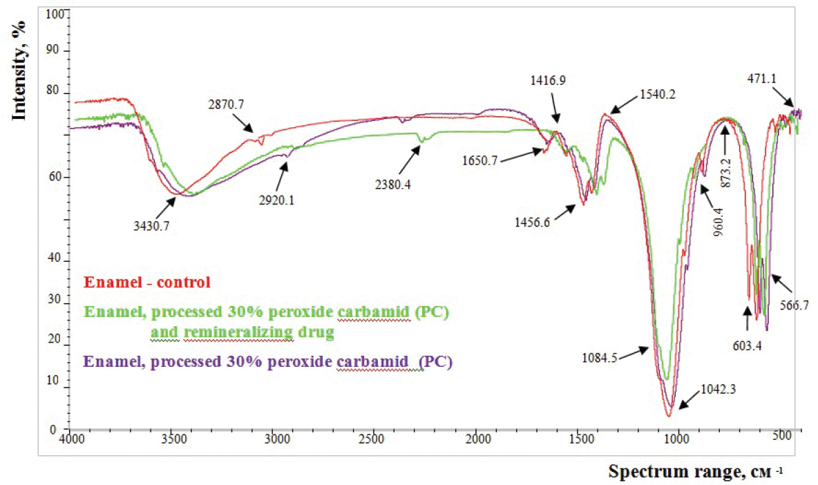
As evidenced by the results of the enamel study, after the application of the drug composition with 30% or 15% of CP (Figs

Analysis of the mineral component of the enamel allowed determining that the parameters of the characteristic band with a maximum at 1040 cm-1 remain the largest. The decrease in the intensity of the valence fluctuations of the PO43- phosphate anion in the spectral region of 1090–1040 cm-1 was noted. No condensed phosphate groups were found that would be characterized by two types of bridge-links P-O-P with fluctuations of 750 cm-1 and 710 cm-1. In our opinion, their absence is due to the greater sensitivity of the bridge links of P-O-P to the influence of oral fluid, in contrast to the stable orthophosphate groups contained in the hydroxyapatite crystal.
In the oscillation region of the connections of the SO32- anion, the absorption bands were determined due to asymmetric flat valence vibrations ν3 with maxima at 1417 cm-1 and 1457 cm-1, as well as unexpressed valence fluctuations ν2 (880–875 cm-1). The parameters of these absorption bands are similar in the control group of samples.
Our obtained results indicate that the absorption band intensity of the hydroxide groups of the short-wave spectrum is reduced, as well as the bandwidths of the valence oscillations of the groups (CH- and NH-) with maxima at 2920 cm-1, 2870 cm-1, 2380 cm-1.
To determine the quantitative changes in the organic and mineral components of the enamel processed by 30% and 15% preparations of dental CP, we carried out the calculation of the optical densities of absorption bands of enamel. The analysis of the mineral enamel matrix has established the stability of the valence grouping parameters in enamel samples processed by different CP concentrations, as well as the lack of changes characterizing the destruction of the organic enamel matrix of the tooth in the Amide I and Amide II regions.
According to the results of the study, after the action of 30% CP, statistically significant changes (p < 0.01) of optical density occurred in the region of the band with a frequency of 3430 cm-1 (water, hydroxide groups), 2920 cm-1 (CH), 2380 cm-1 (НРО42-, СН, NH), as well as a tendency (р > 0.1), in relation to changes in the frequencies of Amide I (1650 cm-1) and Amide II (1540 cm-1).
In the group of enamel samples, a 30% carbamide peroxide preparation determined a statistically significant decrease of 2.17% of the optical density of the band with a frequency of 3430 cm-1 (p < 0.01). Such changes can be assumed to occur due to the ability of urea to form strong hydrogen bonds and to “drag" free water to “itself", that is, towards the surface of the enamel. The water balance of the enamel is restored due to the ability of the centripetal motion of the enamel liquor in the state of the pulp. Taking the above mentioned into consideration, subject to the bleaching of the vital teeth, it can be assumed that changes in the number of hydroxide groups of free and bound water are temporary and reversible.
When comparing the prolonged effect of a bleaching agent of lower concentration (15% CP), the character of the changes in the band of hydroxide groups with a frequency of 3430 cm-1 indicates that the tendency to enamel deamination is preserved (p < 0.05). Compared with the control, the optical density of this band decreased by 1.44%. The figures obtained are summarized in Table

Indices of the optical density of absorption bands of enamel (M ± m), exposed to bleaching agents with different concentrations of urea peroxide.
| Bandwidth absorption (cm -1) | Optical density of absorption bands of enamel (conditional units) | ||
|---|---|---|---|
| Control (n = 17) | The enamel exposed to the medications | ||
| 30% carbamide peroxide (СP) (n = 12) | 15% carbamide peroxide (СP) (n = 14) | ||
| 3430 | 0.138 ± 0.004 | 0.135 ± 0.021** | 0.136 ± 0.018** |
| 2920 | 0.062 ± 0.040 | 0.058 ± 0.06** | 0.061 ± 0.002* |
| 2870 | 0.041 ± 0.003 | 0.039 ± 0.003** | 0.04 ± 0.002* |
| 2380 | 0.006 ± 0.007 | 0.0078 ± 0.002** | 0.066 ± 0.060** |
| 1650 | 0.047 ± 0.006 | 0.046 ± 0.048*** | 0.046 ± 0.021*** |
| 1540 | 0.064 ± 0.012 | 0.063 ± 0.087*** | 0.063 ± 0.046*** |
| 1456 | 0.138 ± 0.002 | 0.138 ± 0.013*** | 0.138 ± 0.025*** |
| 1417 | 0.128 ± 0.002 | 0.128 ± 0.023*** | 0.128 ± 0.044*** |
| 1084 | 0.846 ± 0.012 | 0.845 ± 0.011*** | 0.845 ± 0.016*** |
| 1040 | 1.326 ± 0.015 | 1.323 ± 0.093** | 1.325 ± 0.017** |
| 960 | 0.255 ± 0.003 | 0.255 ± 0.043*** | 0.255 ± 0.062*** |
| 874 | 0.075 ± 0.003 | 0.077 ±0.012* | 0.077 ± 0.014* |
| 603 | 0.432 ± 0.033 | 0.427 ± 0.035** | 0.426 ± 0.073** |
| 566 | 0.437 ± 0.014 | 0.436 ± 0.023* | 0.436 ± 0.013* |
The results indicate that teeth whitening with 15% СP at home, compared to the clinical application of 30% СP, causes a smaller (0.73%) change in the hydrated membranes of enamel prisms. Increased sensitivity of the teeth should be expected to appear during clinical examination in patients due to the processes of dehydration and changes in osmotic pressure of the enamel liquor. Therefore, in the event of hyper-transitions, it is advisable to increase the time interval between sessions of teeth whitening to restore the water balance of the enamel naturally.
Valence oscillations of the amide groups of proteins are localized in the range of 2920–2860 cm-1, 2390–2370 cm-1, 1700–1500 cm-1. In the analysis of the oscillation frequency of 2920 cm-1 and 2870 cm-1, it should be noted that these absorption bands, in addition to the fluctuations of C-H groups, characterize the variations of the hydroxide groups bonded by hydrogen bonds, as well as the R- NH3+, aldehyde, and carboxyl groups. Carboxylic acids and aldehydes should be excluded from consideration since they do not have an absorption characteristic on the spectrum with a frequency of 1700 cm-1.
The analysis of the results of the infrared spectrum of the enamel processed by 30% of the СP product shows a probable reduction in the optical density of absorption bands at maxima of 2920 cm-1 and 2870 cm-1 by 6.8% and 5.1%, according to the control group of samples (p < 0.01). Similar changes were confirmed after tooth brushing with 15% СP: 1.72% and 2.56% (p < 0.05). The obtained results can be assumed to indicate partial destruction of carbon bonds in the range of 2920–2860 cm-1, which is likely to change the dental color in the direction towards lighter colors, due to the transition of absorption of the light beam to the other part of the spectrum.
In addition, attention should be paid to the changes in the region of the spectrum of 2390–2370 cm-1, where the intensity band was reached at a maximum of 2380 cm-1 (НРО42-, C-H- and N-H-grouping). The optical density of this band after the application of 30% of the СP product increased significantly by 30.0%, and after application of 15% of the preparation of СP – by 9.95% (p < 0.01) (Table
To determine the state of the mineral component of bleached teeth, the analysis of the optical densities of absorption bands of orthophosphate-specific bands with maxima of 1085 cm-1, 1040 cm-1, 960 cm-1, 566 cm-1 and carbonates with maxima at 1456 cm-1 and 1417 cm-1, respectively was conducted. The frequencies in the range of 1000–1100 cm-1 are due to the fluctuation of the Н2РО4-, НРО42- and НРО43- ions, and at the frequency of 960 cm-1, the absorption of the РО43-group supplements the absorption of δ (С-Н). Accordingly, it is problematic to determine the changes in the valence oscillations inherent in the РО43- anion, or C-N groups.
After application of 30% or 15% of the CP preparation, no significant decrease in the optical density in the orthophosphate region with absorption band maxima at 1085 cm-1, 1040 cm-1, 960 cm-1 (p > 0.1) (Tab.
In the frequency region of 1456 cm-1 and 1417 cm-1, corresponding to the variation ν1 of the СО32- ion, and also δ C-H, no probable changes in optical absorption density were detected (p>0.1). In the range of 900–850 cm-1 spectrum, after the treatment of the teeth with 15% and 30% СP, an insignificant increase of 2.7% of the optical density of СО32- was determined in comparison with the control group of samples (p > 0.1). Thus, the results of the study indicate that there are no significant changes in the organic or mineral component of the enamel of the tooth, exposed to the action of various concentrations of СP. However, due to the sensitivity of the IRS method to the slightest changes in the nature of valence groups, a tendency has been observed regarding the reduction of soluble forms of phosphates, hydroxide groups, and the nature of ether-phosphorous bonding of enamel. In view of the results obtained, it is advisable to study the effect of remineralizing therapy on the nature of valence groupings of enamel-bleached teeth in order to prevent possible changes in the crystal lattice and organic enamel matrix.
The character of valence groups of bleached enamel after remineralization therapy is as follows. It should be noted that remineralization therapy has taken place since the fifth session of the use of bleaching substances. To do this, at the end of the teeth whitening session, the “Belagel Ca/P" gel preparation was applied on the cleaned and dried surface of the enamel, which was saturated with Ca and P ions for a prolonged time. The IRS results of the enamel (Figs
The study of the spectrograms of the experimental samples did not reveal changes in the number of absorption bands and their localization in comparison with the control group, which indicates the absence of additional inclusions in the chemical structure of the enamel (Table
Indices of the optical density of absorption bands of enamel (M ±m), after treatment with bleaching agents with different CP concentrations and remineralizing therapy.
| Frequency of absorption bands, (см –1) | Optical density of absorption bands of enamel (conditional units) | ||
|---|---|---|---|
| Control (n = 17) | The enamel exposed to the medications | ||
| 30% carbamide peroxide (CP), «Belagel Ca/P» (n = 12) | 15% carbamide peroxide (CP), «Belagel Ca/P» (n = 14) | ||
| 3430 | 0.138 ± 0.004 | 0.143 ± 0.005** | 0.142 ± 0.003** |
| 2920 | 0.062 ± 0.040 | 0.061 ± 0.005* | 0.06 ± 0.004** |
| 2870 | 0.041 ± 0.003 | 0.04 ± 0.024* | 0.04 ± 0.009* |
| 2380 | 0.006 ± 0.007 | 0.0076 ± 0.003** | 0.0075 ± 0.007** |
| 1650 | 0.047 ± 0.006 | 0.046 ± 0.072*** | 0.046 ± 0.061*** |
| 1540 | 0.064 ± 0.012 | 0.063 ± 0.058*** | 0.063 ± 0.092*** |
| 1456 | 0.138 ± 0.002 | 0.139 ± 0.043*** | 0.138 ± 0.074*** |
| 1417 | 0.128 ± 0.002 | 0.127 ± 0.013*** | 0.127 ± 0.042*** |
| 1084 | 0.846 ± 0.012 | 0.845 ± 0.01*** | 0.845 ± 0.013*** |
| 1040 | 1.326 ± 0.015 | 1.329 ± 0.014** | 1.328 ± 0.014** |
| 960 | 0.255 ± 0.003 | 0.254 ± 0.004*** | 0.255 ± 0.003*** |
| 874 | 0.075 ± 0.003 | 0.078 ± 0.003** | 0.079 ± 0.003** |
| 603 | 0.432 ± 0.033 | 0.429 ± 0.034** | 0.427 ± 0.038** |
| 566 | 0.437 ± 0.014 | 0.438 ± 0.037*** | 0.437 ± 0.052*** |
Further analysis of the absorption strips of bleached enamel samples and the Belagel Ca/P was performed at 2920–2860 cm-1 (p < 0.01). The obtained results indicate that the use of remineralizing agent does not affect the process of destruction of the carbon bonds of CH- and NH-groups and the removal of the restructured organic component of the enamel (probably this process relates to the chromoform of the pigments) when bleaching the teeth. A probable reduction of the absorption band with a maximum at 603 cm-1 was determined, and an increase in the optical density index with a maximum at 874 cm-1 and 1040 cm-1, indicating a process of saturation of enamel with НРО42- ions (p < 0.05).
The use of remineralizing therapy leads to a possible increase in the optical density of the band with a maximum at 3430 cm-1 (p < 0.01) in the case of teeth whitening in clinical conditions by 5.79%, and at home – by 3.74% – according to indicators of bleached teeth.
Taking into account the obtained results, it can be assumed that the remineralization capacity of the oral fluid and the pulp, the state of hydration membranes, osmotic pressure of the enamel liquor is restored in the enamel due to the compensatory processes caused by the action of the Belagel Ca/P. Because of these activities, the processes of ion exchange are improved. Therefore, the use of remineralizing therapy in a clinical study of bleaching agents can lead to the disappearance of an unpleasant sensation during tooth whitening due to changes in the pulp.
Conclusions
Thus, the obtained results of the study indicate that the process of discoloration of teeth is accompanied by changes in carbon bonds of СН- and NH-groups, a decrease in the number of OH-groups and dissolved forms of phosphates. When applying medicinal compositions, in accordance with the elaborated methods of bleaching the vital teeth, no significant changes in the enamel are revealed, which would indicate the destruction of the hydroxyapatite crystals or its organic matrix. To seal the crystal lattice of enamel, the use of remineralizing therapy with drugs containing Ca and R ions is recommended.
References
- Bell RJ (1975) Introduction to Fourier – Spectroscopy. “World": M.,160 pp.
- Ciurczak EW, Drennen JК (2002) Pharmaceutical and Medical Applications of Near-infrared Spectroscopy (Practical Spectroscopy). Marcel Decker, New York, 38 pp. https://doi.org/10.1201/9780203910153
- Ferraro JR, Krishnan K (1990) Practical Fourier Transform Infrared Spectroscopy. Industrial and Laboratory Chemical Analysis. Academic Press, New York, 548 pp.
- Gordetsov AS (2010) Infrared spectroscopy of biological fluids and tissues. Modern Technologies in Medicine 1: 84–98.
- Hrynovets І, Mahlovanyy A, Deneha I, Ripetska O (2016) Application of different medicinal forms in dental practice Gorlice, (RP), 105 pp.
- Kargapolov AV, Zubareva GM, Mikin VM (2009) Infrared spectrometry in the study of oral fluid for diagnostic purposes. Dentistry 5: 7–10.
- Kazarina LN, Rounova OA, Vdovina LV (2014) Infrared spectroscopy as a method for early diagnosis of caries. Modern Problems of Science and Education [Online]: 6. http://www.science-education.ru/120-16287
- Kazarina LN, Smetanina OA, Gordetsov AS, Krasnikova OV (2016) Infrared spectroscopy of oral fluid as a method for the early diagnosis of inflammatory periodontal diseases in children. Modern Problems of Science and Education [Online]: 6. https://www.science-education.ru/ru/article/view?id=25848 [Accessed on: 15th December 2016]
- Mathur KK, Tatuіn SA, Keilman RM (2003) Carbonated apatite and hydroxide apatite reconstruction. Archives of Facial Plastic Surgery 5: 379–383. https://doi.org/10.1001/archfaci.5.5.379
- Prech E, Buhlmann F, Affolter K (2006) Determination of the structure of organic compounds. Tables of spectral data. M.: Mir; BINOMIAL. Laboratory of knowledge, 438 pp.
- Sidorchuk MV (2012) Applied Infrared Spectroscopy. In: Kendall D (Ed. ) PVD «Tverdynya», Lutsk, 128 pp.
- Smith BC (1996) Fundamentals of Fourier Transform Infrared Spectroscopy. CRC Press: Boca Raton: FL, 203 pp.
- Stuart BH (2004) Infrared Spectroscopy: Fundamentals and Applications. Wiley, 244 pp. https://doi.org/10.1002/0470011149
- Workman J, Weyer L (2008) Practical Guide to Interpretive Near-Infrared Spectroscopy. CRS Press Taylor & Francis Group: Boca Raton: FL, 108 pp. https://doi.org/10.1201/9781420018318
- Zubachyk VM, Kononenko VV, Sinitsya VV (2001) Mechanism of discoloration of teeth. Herald in Dentistry 2: 17–18.
- Zubachyk VM, Kononenko VV, Sinitsya VV (2003) Patent 55071А Ukraine, IPC (2002.06) A61K6/02. Method of teeth whitening. [published 17.03.2003. – Bull No. 3.]