Research Article |
|
Corresponding author: Kurnia Agustini ( kurn005@brin.go.id ) Corresponding author: Frangky Sangande ( frangky.sangande@gmail.com ) Academic editor: Ivan Dimitrov
© 2024 Kurnia Agustini, Frangky Sangande, Nuralih Nuralih, Armansyah Maulana Harahap, Sri Ningsih, Anton Bahtiar.
This is an open access article distributed under the terms of the Creative Commons Attribution License (CC BY 4.0), which permits unrestricted use, distribution, and reproduction in any medium, provided the original author and source are credited.
Citation:
Agustini K, Sangande F, Nuralih N, Harahap AM, Ningsih S, Bahtiar A (2024) Molecular mechanism elucidation of Ocimum basilicum as anticancer using system bioinformatic approach supported by in vitro assay. Pharmacia 71: 1-12. https://doi.org/10.3897/pharmacia.71.e127395
|
Abstract
Breast cancer (BC) is a multifactorial disease involving many pathways and target molecules. Multi-target therapy through multi-compound herbal medicines is an alternative strategy to treat BC. In the present study, we elucidate the molecular mechanism of Ocimum basilicum (OB) as an anticancer agent using system bioinformatic approach and investigate its cytotoxic effect against MCF-7 cells. We performed network pharmacology (NP) and molecular docking studies to provide scientific information regarding the underlying anti-BC mechanism of OB. Based on topology parameters obtained from protein-protein interaction (PPI), we identified six potential targets that play a significant role in the network including SRC, PI3KCA, EGFR, ESR1, AKT1, and MAPK1. Furthermore, consensus docking suggested rutin, quercetin-3-O-diglucoside, and kaempferol-3-O-β-D-rutinoside as the potential compounds of OB. Moreover, Kyoto Encyclopedia of Genes and Genomes (KEGG) analysis demonstrated that the cytotoxic effect of OB might be related to the modulation of several pathways such as PI3K-Akt, VEGF, and HIF-1, breast cancer, and estrogen signaling pathways. The in vitro assay revealed that various extracts of OB demonstrated cytotoxic effects against MCF-7 with IC50 = 231 µg/mL (OB ethanolic extract), 408 µg/mL (OB methanolic extract), 479 µg/mL (OB ethyl acetate extract), 1887 µg/mL (OB n-hexanoic extract) and 767 µg/mL (OB butanolic extract) respectively.
Keywords
Ocimum basilicum, MCF-7 cells, Cytotoxicity, Network Pharmacology, Molecular Docking
Introduction
Breast cancer (BC) is one of the most common cancers diagnosed in women worldwide, and its occurrence exceeded lung cancer for the first time in 2020 (
The underlying molecular mechanisms of BC are complex involving dysregulation of various signaling pathways with their respective target molecules (
Based on current data, of 250,000 plant species found in the kingdom Plantae, only 10% of species have been explored for their anticancer activity (
As a multifactorial disease, molecular mechanism elucidation of BC is challenging at the cellular level, particularly when using multi-compound substances (e.g. extract) since the biological activity is an accumulation of multi-target and multi-compound interactions. Therefore, it is important to consider all possible targets modulated by the compounds. To address this, network pharmacology (NP) offers a systematic and comprehensive approach to analyzing the molecular mechanism of extract materials (
Materials and methods
Collection of active compounds of OB and their potential targets
The active compounds of OB were collected from Dr. Duke’s Phytochemical and Ethnobotanical Databases (https://phytochem.nal.usda.gov/) by focusing on leaf part and their potential targets with probability ≥ 0.1 were predicted using SwissTargetPrediction (http://www.swisstargetprediction.ch/). The target obtained in this step was termed “compound-related target”.
ERPBC-related targets of OB identification
To collect disease-related targets of OB, we used two databases: GeneCards and DisGeNet. Due to ~75% of all BC cases being diagnosed as estrogen receptor alpha-positive (ERα+) and MCF-7 representing this BC subtype (
Protein-protein interaction (PPI) construction and KEGG analysis
The STRING database (https://string-db.org/) on Homo sapiens with a high confidence score (0.7) was used to construct the PPI network by submitting ERPBC-related targets of OB. Unconnected targets were deleted and the remaining targets were submitted to ShyniGO 0.77 (http://bioinformatics.sdstate.edu/go/) to analyze KEGG enrichment at the p-value of ≤ 0.05. This step resulted in the top 20 enriched pathways ranked by false discovery rate (FDR).
Potential targets identification
The result of the PPI construction above was then submitted to Cytoscape 3.9.3 and the topological features for each target include local average connectivity (LAC), closeness centrality (CC), eigenvector centrality (EC), network centrality (NC), and degree centrality (DC) were analyzed using CytoNCA. Targets with topological scores greater than the average score were considered potential targets.
Potential compounds identification
Molecular docking was used to identify the virtual hits for every potential target by screening all compounds having targets with probability ≥ 0.1 in step 2.4. The 3D structure of these compounds was retrieved from PubChem. The structures of the main targets: SRC (PDB: 2BDJ), PI3KCA (PDB: 5DXT), EGFR (PDB: 1M17), ESR1 (PDB: 3ERT), AKT1 (PDB: 4GV1), MAPK1 (PDB: 3I5Z) were downloaded from RCSB. DOCK6 and Vina were used for docking simulations using a consensus method and the protocols were developed based on a previous study (
In DOCK6, targets and ligands were added charge using AM1-BBC method. A probe radius of 1.4 Å was used to generate the molecular surface. The active site of a target was determined using spheres within 8 Å of the native ligand which is surrounded by a box with a margin of 5 Å.
In Vina, the targets were prepared by adding polar hydrogens followed by Kollman charges. Meanwhile, Gestaiger charges were used in ligand preparation. A grid box centered on a native ligand position at a spacing of 1 Å was applied to generate the center coordinate. The size box of SRC, PI3KCA, ESR1, AKT1, and MAPK1 was set to 22 × 22 × 22 Å, while the size of 20 × 18 × 18 Å was used in EGFR.
The 10 conformations per ligand generated from the two docking tools were compared with each other, and compounds having duplet conformation in DOCK6 and Vina were calculated for their consensus score by averaging the binding energy (kcal/mol) of these conformations in each tool. Compounds getting the best consensus score were defined as virtual hits.
Sample preparation
OB was collected in July 2023 from Bogor, West Java, Indonesia. The leaves were dried, grinded, and extracted with various solvents such as methanol, ethanol, ethyl acetate, butanol, and n-hexane. Then a vacuum rotating evaporator was used to dry the filtrate.
Cell culture
We used MCF-7 cell lines from Human Breast Adenocarcinoma, that were obtained from the cell culture collection at the Laboratory for Development of Industrial Agro and Biomedical Technology (LAPTIAB) PUSPIPTEK, National Research and Innovation Agency (BRIN), Serpong, South Tangerang, Indonesia. MCF-7 cells were cultivated in medium RPMI 1640 (Gibco Life Technologies) supplemented with 10% heat-inactivated Fetal Bovine Serum (FBS, Gibco Life Technologies), phenol red, 100 U/ml penicillin, 0.1 mg/ml streptomycin, 2 mM glutamine and 1 mM sodium pyruvate. Cells were maintained in 75 cm2 flasks at 37 °C, 5% CO2, and 95% humidity.
Cytotoxic assay
In 96-well plates, 10,000 cells/well were platted using medium RPMI with phenol red containing 10% Fetal Bovine Serum (FBS), 0.1 mg/ml streptomycin, 100 U/ml penicillin and 1mM sodium pyruvate. Cells were incubated for 24 hours at 37 °C, 5% CO2 and in a 95% humidified atmosphere, then the medium was changed with samples in growth medium in six variation concentrations and incubated for another 24 hours at 37 °C, 5% CO2, and in a 95% humidified atmosphere. After that, Phosphate Buffer Saline (PBS) solution was added to wash the cell. To identify the living cells, we added the solution of MTT (3-(4,5-Dimethylthiazol-2-yl)-2,5-Diphenyltetrazolium) in medium followed by incubation for 4 hours at 37 °C, 5% CO2, and in a 95% humidified atmosphere. During this incubation, the crystal of formazan blue will be formed. At last, we added the Sodium Dodecyl Sulphate (SDS) into every well to stop the reaction and leave the plate in a dark place for 12 hours (overnight). The intensity of the color formed was measured by ELISA reader at 570 nm. Data were analyzed with Probit Analysis to obtain the IC50. (
Results
Active compounds of OB and their target prediction
A total of 44 compounds contained in OB leaves were retrieved from the database, and among them, only 28 compounds were found to have targets at probability ≥ 0.1 by SwissTargetPrediction. After analyzing, we identified 321 compound-related targets at this step.
ERPBC-related targets of OB
From GeneCards and DisGeNet, we collected 756 ERPBC-related targets. After merging these ERPBC-related targets with compound-related targets, we found 67 common targets in the intersection area (Fig.

PPI and potential target
For further analysis, the PPI of the common targets was constructed using STRING. Of 67 targets, only 58 were connected to each other, and their interactions are shown in Fig.

| Target | Topology Parameter | |||||
|---|---|---|---|---|---|---|
| Degree | Eigenvector | LAC | Betweenness | Closeness | Network | |
| SRC | 21 | 0.4 | 5.0 | 638.6 | 0.5 | 15.5 |
| PIK3CA | 17 | 0.4 | 5.3 | 243.3 | 0.5 | 12.5 |
| EGFR | 13 | 0.3 | 4.5 | 212.1 | 0.4 | 7.1 |
| ESR1 | 14 | 0.3 | 3.4 | 1267.0 | 0.5 | 7.0 |
| AKT1 | 14 | 0.2 | 2.6 | 426.3 | 0.5 | 6.3 |
| MAPK1 | 16 | 0.2 | 2.4 | 542.9 | 0.5 | 5.6 |
| Cutoff | 5.3 | 0.1 | 1.7 | 118.3 | 0.3 | 2.9 |
KEGG analysis
After submitting the 58 targets mentioned above to ShinyGO, 20 enriched pathways were obtained. Interestingly, in addition to PI3K-Akt, VEGF, and HIF-1 signaling pathways, breast cancer and estrogen signaling pathways which are closely related to ERPBC were also detected in this result (Fig.

Potential compounds
Based on NP studies, six potential targets might be modulated by OB for ERPBC treatment. To verify whether active compounds of OB can bind to these targets, we performed molecular docking simulations. Before simulations, the docking protocol of DOCK6 and Vina was validated by redocking the co-crystals to their corresponding target. Table
| Target | DOCK6 | Vina |
|---|---|---|
| SRC |

|
|
| PI3KCA |

|

|
| EGFR |

|

|
| AKT1 |

|
|
| MAPK1 |

|

|
Docking results of 28 active compounds of OB revealed that there were different hits between DOCK6 and Vina. For instance, kaempferol-3-O-β-D-rutinoside and quercetin-3-O-diglucoside were compounds with the most negative score (-10.5 kcal/mol) on SRC based on Vina (Table
Docking score (kcal/mol) of duplet conformation on main targets using Vina.
| No | Compound | Vina score | |||||
|---|---|---|---|---|---|---|---|
| SRC | PI3KCA | EGFR | ESR1 | AKT1 | MAPK1 | ||
| 1 | Rosmarinic acid | – | -7.5 | – | -7.3 | -8.1 | – |
| 2 | Cinnamic acid | – | -6.3 | – | – | -6.4 | – |
| 3 | Ferulic acid | -6.6 | -6.3 | -6.0 | – | -5.8 | -6.1 |
| 4 | Narigenin | -9.0 | -8.9 | -8.6 | -8.7 | -7.8 | -8.3 |
| 5 | Aesculetin | -6.6 | -6.5 | -6.3 | – | -6.7 | -6.4 |
| 6 | Aesculin | -8.2 | -7.6 | -9.1 | – | -7.4 | -8.2 |
| 7 | Benzyl acetate | -5.8 | -5.9 | -5.3 | – | – | – |
| 8 | Caffeic acid | -6.6 | -6.3 | – | – | – | – |
| 9 | Caffeic acid n-butyl ester | -6.6 | -7.1 | – | -6.6 | -6.2 | – |
| 10 | Campesterol | – | – | -8.7 | – | -7.7 | -8.0 |
| 11 | Cinnamic acid methyl ester | – | – | -5.2 | – | -5.7 | – |
| 12 | Eriodictyol | -9.4 | -9.2 | -8.6 | -8.0 | -7.9 | -8.7 |
| 13 | Eriodictyol-7-O-β-D-glucoside | -9.8 | -10.2 | -9.7 | -7.7 | -9.3 | -9.6 |
| 14 | Estragole | -5.6 | -5.8 | – | – | -5.8 | -5.5 |
| 15 | Eugenol | -6.0 | -5.5 | -5.4 | – | -6.5 | – |
| 16 | Isoquercitrin | -10.3 | -9.0 | -8.2 | -7.5 | – | – |
| 17 | Kaempferol | -9.6 | -8.5 | -8.7 | -8.2 | -7.4 | – |
| 18 | Kaempferol-3-O-β-D-rutinoside | -10.5 | -9.4 | -8.6 | – | -9.0 | -9.2 |
| 19 | p-coumaric acid | – | -6.7 | -6.0 | – | -6.6 | -6.3 |
| 20 | Quercetin | -9.7 | -8.5 | -8.9 | -7.9 | -7.7 | -8.9 |
| 21 | Quercetin-3-O-diglucoside | -10.5 | -9.2 | -9.4 | -7.7 | -8.9 | – |
| 22 | Rutin | -9.9 | -9.8 | -9.2 | – | -9.1 | – |
| 23 | Salicylic acid-2-β-D-glucoside | -7.3 | -6.6 | -8.6 | -6.6 | -6.7 | -6.7 |
| 24 | Thymol | -6.4 | -6.0 | -5.8 | – | -6.3 | -6.1 |
| 25 | Ursolic acid | – | -6.7 | -8.5 | -9.1 | -7.0 | |
| 26 | Vanillic acid-4-β-D-glucoside | -7.1 | -6.8 | -7.9 | -6.2 | -7.8 | -7.2 |
| 27 | Vicenin 2 | -9.9 | -9.1 | -9.1 | – | – | -8.3 |
| 28 | Xanthomicrol | – | – | – | – | – | – |
| 29 | Co-crystal of SRC | -10.1 | |||||
| 30 | Co-crystal of PI3KCA | -9.1 | |||||
| 31 | Co-crystal of EGFR | -7.4 | |||||
| 32 | Co-crystal of ESR1 | -9.9 | |||||
| 33 | Co-crystal of AKT1 | -8.5 | |||||
| 34 | Co-crystal of MAPK1 | -10.5 | |||||
Docking score (kcal/mol) of duplet conformation on main targets using DOCK6.
| No | Compound | DOCK6 score | |||||
|---|---|---|---|---|---|---|---|
| SRC | PI3KCA | EGFR | ESR1 | AKT1 | MAPK1 | ||
| 1 | Rosmarinic acid | – | -58.6 | – | -62.2 | -62.7 | – |
| 2 | Cinnamic acid | – | -29.9 | – | – | -32.2 | – |
| 3 | Ferulic acid | -40.9 | -35.8 | -35.6 | – | -38.6 | -38.0 |
| 4 | Narigenin | -48.4 | -46.4 | -46.3 | -47.4 | -46.8 | -47.2 |
| 5 | Aesculetin | -32.3 | -33.2 | -33.0 | – | -34.8 | -33.9 |
| 6 | Aesculin | -51.9 | -51.7 | -52.7 | – | -51.9 | -54.4 |
| 7 | Benzyl acetate | -33.7 | -29.9 | -31.4 | – | – | – |
| 8 | Caffeic acid | -37.2 | -35.1 | – | – | – | – |
| 9 | Caffeic acid n-butyl ester | -47.5 | -46.4 | – | -47.4 | -47.4 | – |
| 10 | Campesterol | – | – | -51.6 | – | -57.1 | -48.4 |
| 11 | Cinnamic acid methyl ester | – | – | -34.1 | – | -32.2 | – |
| 12 | Eriodictyol | -50.4 | -49.1 | 49.5 | -46.7 | -50.7 | -47.7 |
| 13 | Eriodictyol-7-O-β-D-glucoside | -67.3 | -67.4 | -61.8 | -56.9 | -68.3 | -65.7 |
| 14 | Estragole | -32.9 | -29.5 | – | – | -32.1 | -31.4 |
| 15 | Eugenol | -34.8 | -32.6 | -32.9 | – | -34.7 | – |
| 16 | Isoquercitrin | -62.7 | -61.3 | -55.5 | -54.2 | – | – |
| 17 | Kaempferol | -50.0 | -48.5 | -47.2 | -46.8 | -46.6 | – |
| 18 | Kaempferol-3-O-β-D-rutinoside | -80.7 | -75.8 | -72.0 | – | -75.5 | -73.4 |
| 19 | p-coumaric acid | – | -33.2 | -32.4 | – | -34.6 | -34.2 |
| 20 | Quercetin | -52.4 | -49.9 | -49.9 | -46.8 | -45.8 | -48.1 |
| 21 | Quercetin-3-O-diglucoside | -80.5 | -77.1 | -62.9 | -70.3 | -83.6 | – |
| 22 | Rutin | -82.7 | -75.5 | -70.9 | – | -79.9 | – |
| 23 | Salicylic acid-2-β-D-glucoside | -49.2 | -46.2 | -49.6 | -51.9 | -47.6 | -51.8 |
| 24 | Thymol | -31.3 | -30.3 | -29.8 | – | -30.7 | -30.6 |
| 25 | Ursolic acid | – | -44.4 | -42.0 | -52.6 | -52.3 | -41.3 |
| 26 | Vanillic acid-4- β-D-glucoside | -50.9 | -50.1 | -52.2 | -53.0 | -53.5 | -54.2 |
| 27 | Vicenin 2 | -78.3 | 67.4 | -68.9 | – | – | -62.1 |
| 28 | Xanthomicrol | – | – | – | – | – | – |
| 29 | Co-crystal of SRC | -85.4 | |||||
| 30 | Co-crystal of PI3KCA | -73.2 | |||||
| 31 | Co-crystal of EGFR | -64.3 | |||||
| 32 | Co-crystal of ESR1 | -88.3 | |||||
| 33 | Co-crystal of AKT1 | -95.0 | |||||
| 34 | Co-crystal of MAPK1 | -88.0 | |||||
As shown in Table

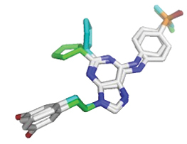
H-bond profiles of co-crystal on SRC (A1); rutin on SRC (A2); co-crystal on PI3KCA (B1); quercetin-3-O-diglucoside on PI3KCA (B2); co-crystal on EGFR (C1); kaempferol-3-O-β-D-rutinoside on EGFR (C2). Dashed green lines represent hydrogen bonds. Yellow sticks represent the residues of targets. Brown sticks represent the ligands.

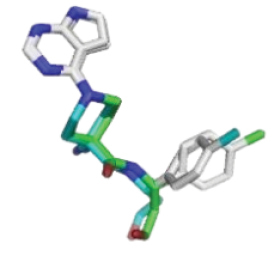
H-bond profiles of co-crystal on ESR1 (A1); quercetin-3-O-diglucoside on ESR1 (A2); co-crystal on AKT1 (B1); quercetin-3-O-diglucoside on AKT1 (B2); co-crystal on MAPK1 (C1); kaempferol-3-O-β-D-rutinoside on MAPK1 (C2). Dashed green lines represent hydrogen bonds. Yellow sticks represent the residues of targets. Brown sticks represent the ligands.
| No | Compound | Consensus score | |||||
|---|---|---|---|---|---|---|---|
| SRC | PI3KCA | EGFR | ESR1 | AKT1 | MAPK1 | ||
| 1 | Rosmarinic acid | – | -33.1 | – | -34.8 | -35.4 | – |
| 2 | Cinnamic acid | – | -18.1 | – | – | -19.3 | – |
| 3 | Ferulic acid | -23.8 | -21.1 | -20.8 | – | -22.2 | -22.1 |
| 4 | Narigenin | -28.7 | -27.7 | -27.5 | -28.1 | -27.3 | -27.8 |
| 5 | Aesculetin | -19.5 | -19.9 | -19.7 | – | -20.8 | -20.2 |
| 6 | Aesculin | -30.1 | -29.7 | -30.9 | – | -29.7 | -31.3 |
| 7 | Benzyl acetate | -19.8 | -17.9 | -18.4 | – | – | – |
| 8 | Caffeic acid | -21.9 | -20.7 | – | – | – | – |
| 9 | Caffeic acid n-butyl ester | -27.1 | -26.8 | – | -27.0 | -26.8 | – |
| 10 | Campesterol | – | – | -30.2 | – | -32.4 | -28.2 |
| 11 | Cinnamic acid methyl ester | – | – | -19.7 | – | -19.0 | – |
| 12 | Eriodictyol | -29.9 | -29.2 | -20.5 | -27.4 | -29.3 | -28.2 |
| 13 | Eriodictyol-7-O-β-D-glucoside | -38.6 | -38.8 | -35.8 | -32.3 | -38.8 | -37.7 |
| 14 | Estragole | -19.3 | -17.7 | – | – | -19.0 | -18.5 |
| 15 | Eugenol | -20.4 | -19.1 | -19.2 | – | -20.6 | – |
| 16 | Isoquercitrin | -36.5 | -35.2 | -31.9 | -30.9 | – | – |
| 17 | Kaempferol | -29.8 | -28.5 | -28.0 | -27.5 | -27.0 | – |
| 18 | Kaempferol-3-O-β-D-rutinoside | -45.6 | -42.6 | -40.3 | – | -42.3 | -41.3 |
| 19 | p-coumaric acid | – | -20.0 | -19.2 | – | -20.6 | -20.3 |
| 20 | Quercetin | -31.1 | -29.2 | -29.4 | -27.4 | -26.8 | -28.5 |
| 21 | Quercetin-3-O-diglucoside | -45.5 | -43.2 | -36.2 | -39.0 | -46.3 | – |
| 22 | Rutin | -46.3 | -42.7 | -40.1 | – | -44.5 | – |
| 23 | Salicylic acid-2-β-D-glucoside | -28.3 | -26.4 | -29.1 | -29.3 | -27.2 | -29.3 |
| 24 | Thymol | -18.9 | -18.2 | -17.8 | – | -18.5 | -18.4 |
| 25 | Ursolic acid | – | -25.6 | -25.3 | -26.3 | -30.7 | -24.2 |
| 26 | Vanillic acid-4-β-D-glucoside | -29.0 | -28.5 | -30.1 | -29.6 | -30.7 | -30.7 |
| 27 | Vicenin 2 | -44.1 | 29.2 | -39.0 | – | – | -35.2 |
| 28 | Xanthomicrol | – | – | – | – | – | – |
| 29 | Co-crystal of SRC | -47.8 | |||||
| 30 | Co-crystal of PI3KCA | -41.2 | |||||
| 31 | Co-crystal of EGFR | -35.9 | |||||
| 32 | Co-crystal of ESR1 | -49.1 | |||||
| 33 | Co-crystal of AKT1 | -51.8 | |||||
| 34 | Co-crystal of MAPK1 | -49.3 | |||||
Influence of OB extracts on viability of MCF-7 cells
From cell assay, we found that ethanolic extract has a better cytotoxic effect on MCF-7 cells than the extract with other solvents (Fig.

Discussion
OB is a spicy plant commonly used in traditional medicine for treating several conditions such as dysentery, flatulence, colds, and nausea (
Our docking simulation revealed that almost all compounds were able to bind to these main targets simultaneously, indicating OB has a multitarget and synergic effect, thus increasing their anticancer activity. Based on the consensus score (Table
On PI3KCA, the score of this compound was better than the co-crystal ligand. It formed H-bonds with Ser773, Ser774, and Asp933. Asp933 was reported as the residue that most frequently forms an H-bond with many inhibitors (
Kaempferol-3-O-β-D-rutinoside, on the other hand, was the best-scored compound on EGFR and MAPK1. This compound binds to EGFR by forming H-bonds with Asp766 and Met769, while on MAPK1, it formed H-bonds with Met106, Lys115, and Ser151. Meanwhile, rutin showed the most negative score on SRC and formed H-bonds with Ala293, Lys295, Met341, and Asn391. Met769, Met106, and Met341 are the key residues at the hinge region of EGFR, MAPK1, and SRC, respectively. These targets are members of kinase protein, and it has been reported that H-bonds interaction with the kinase hinge is usually indispensable for potent inhibition (
In this study, we also investigated the potential anticancer activity of some extracts of OB on MCF-7 and found that they have cytotoxic effects with IC50 = 231 µg/mL (ethanolic extract), 408 µg/mL (methanolic extract), 479 µg/mL (ethyl acetate extract), 1887 µg/mL (n-hexanoic extract) and 767 µg/mL (buthanolic extract). MCF-7 is one of BC cell cultures with estrogen receptors. Ethanolic extract provides better cytotoxic activity compared to other extracts. It seems that the compounds responsible for providing cytotoxic activity are absorbed in the ethanol solvent.
Several studies have tested the cytotoxic effect of OB. However, they used different parts (aerial part, seed, essential oil) of OB and different solvents. Moreover, the molecular mechanisms were not characterized (
According to National Cancer Institute (NCI) and Geran protocol, the cytotoxic effect of a substance is categorized as strong (IC50< 20 µg/mL), moderate (IC50 = 21–200 µg/mL), weak (IC50 = 201–500 µg/mL), and non-cytotoxic (IC50 >501 µg/mL) (
Conclusion
Although OB has a weak cytotoxic effect on MCF-7, it showed a good potential for development as an alternative anticancer agent after optimization, such as through the fractionation process. NP studies suggested that OB has cytotoxic activity possibly by modulating several pathways including breast cancer, estrogen, PI3K-Akt, VEGF, and HIF-1 signaling pathways, and targeting six main targets: SRC, PI3KCA, EGFR, ESR1, AKT1, and MAPK1. Meanwhile, three compounds: rutin, quercetin-3-O-diglucoside, and kaempferol-3-O-β-D-rutinoside were considered as the potential compounds of OB. Thus, the cytotoxic activity of OB might partly be due to thebiological activity of these compounds. For the next studies, an in vitro assay against the six main targets is needed to validate this hypothesized molecular mechanism of OB. Moreover, fractionating the crude extract by focusing on the potential compounds might be a strategy to improve the cytotoxic activity.
Conflicts of Interest
There is no conflict of interest between any of the authors. The manuscript hasn’t been released yet, nor has it been offered for publication anywhere. The manuscript’s publishing has the unanimous approval of all authors.
Acknowledgements
This work was funding by Research Grant 2023 from Health Research Organization, National Research and Innovation Agency (BRIN), Indonesia.
References
- Agustini K, Kusumaningrum S, Firdayani, Sulfanti A, Wink M (2023) Estrogenic activity of Bryonia dioica Jacq. through in silico and in vitro studies on pS2 gene expression in the breast cancer cell line MCF-7. Pharmacia 70(4): 951–958. https://doi.org/10.3897/pharmacia.70.e103478
- Al-Ali KH, El-Beshbishy HA, El-Badry AA, Alkhalaf M (2013) Cytotoxic activity of methanolic extract of Mentha longifolia and Ocimum basilicum against human breast cancer. Pakistan Journal of Biological Sciences 16(23): 1744–1750. https://doi.org/10.3923/pjbs.2013.1744.1750
- Aminian AR, Mohebbati R, Boskabady MH (2022) The effect of Ocimum basilicum L. and its main ingredients on respiratory disorders: An experimental, preclinical, and clinical review. Frontiers in Pharmacology 12: 805391. https://doi.org/10.3389/fphar.2021.805391
- Arnold M, Morgan E, Rumgay H, Mafra A, Singh D, Laversanne M, Vignat J, Gralow JR, Cardoso F, Siesling S, Soerjomataram I (2022) Current and future burden of breast cancer: Global statistics for 2020 and 2040. Breast 66: 15–23. https://doi.org/10.1016/j.breast.2022.08.010
- Arshad Qamar K, Dar A, Siddiqui BS, Kabir N, Aslam H, Ahmed S, Erum S, Habib S, Begum S (2010) Anticancer activity of Ocimum basilicum and the effect of ursolic acid on the cytoskeleton of MCF-7 human breast cancer cells. Letters in Drug Design & Discovery 7(10): 726–736. https://doi.org/10.2174/1570180811007010726
- Baktiar Laskar Y, Meitei Lourembam R, Behari Mazumder P (2020) Herbal remedies for breast cancer prevention and treatment, IntechOpen. https://doi.org/10.5772/intechopen.89669
- Bolognesi ML, Rossi M (2020) Multitarget Drug Discovery. Encyclopedia of Life Sciences 1–7. https://doi.org/10.1002/9780470015902.A0028845
- Charles-Okhe O, Odeniyi MA, Fakeye TO, Ogbole OO, Akinleye TE, Adeniji AJ (2022) Cytotoxic activity of crude extracts and fractions of African peach (nauclea latifolia smith) stem bark on two cancer cell lines. Phytomedicine Plus 2(1): 1–9. https://doi.org/10.1016/j.phyplu.2021.100212
- de Diesbach MT, Cominelli A, N’Kuli F, Tyteca D, Courtoy PJ (2010) Acute ligand-independent Src activation mimics low EGF-induced EGFR surface signalling and redistribution into recycling endosomes. Experimental Cell Research 316(19): 3239–3253. https://doi.org/10.1016/j.yexcr.2010.09.001
- Eanes L, Patel YM (2016) Inhibition of the MAPK pathway alone is insufficient to account for all of the cytotoxic effects of naringenin in MCF-7 breast cancer cells. Biochimie Open 3: 64–71. https://doi.org/10.1016/j.biopen.2016.09.004
- Eid AM, Jaradat N, Shraim N, Hawash M, Issa L, Shakhsher M, Nawahda N, Hanbali A, Barahmeh N, Taha B, Mousa A (2023) Assessment of anticancer, antimicrobial, antidiabetic, anti-obesity and antioxidant activity of Ocimum Basilicum seeds essential oil from Palestine. BMC Complementary Medicine and Therapies 23(1): 221. https://doi.org/10.1186/s12906-023-04058-w
- Feng Y, Spezia M, Huang S, Yuan C, Zeng Z, Zhang L, Ji X, Liu W, Huang B, Luo W, Liu B, Lei Y, Du S, Vuppalapati A, Luu HH, Haydon RC, He TC, Ren G (2018) Breast cancer development and progression: Risk factors, cancer stem cells, signaling pathways, genomics, and molecular pathogenesis. Genes & Diseases 5(2): 77–106. https://doi.org/10.1016/j.gendis.2018.05.001
- Giordano S, Petrelli A (2008) From single- to multi-target drugs in cancer therapy: When aspecificity becomes an advantage. Current Medicinal Chemistry 15(5): 422–432. https://doi.org/10.2174/092986708783503212
- Houston DR, Walkinshaw MD (2013) Consensus docking: improving the reliability of docking in a virtual screening context. Journal of Chemical Information and Modeling 53(2): 384–390. https://doi.org/10.1021/ci300399w
- Irwin ME, Bohin N, Boerner JL (2011) Src family kinases mediate epidermal growth factor receptor signaling from lipid rafts in breast cancer cells. Cancer Biology & therapy 12(8): 718–726. https://doi.org/10.4161/cbt.12.8.16907
- Kim JH, Lee MH, Kim BJ, Kim JH, Han SJ, Kim HY, Stallcup MR (2005) Role of aspartate 351 in transactivation and active conformation of estrogen receptor α. Journal of Molecular Endocrinology 35(3): 449–464. https://doi.org/10.1677/jme.1.01846
- Li J, Zhou N, Liu W, Li J, Feng Y, Wang X, Wu C, Bao J (2016) Discover natural compounds as potential phosphodiesterase-4B inhibitors via computational approaches. Journal of Biomolecular Structure & Dynamics 34(5): 1101–1112. https://doi.org/10.1080/07391102.2015.1070749
- Mahajan P, Wadhwa B, Barik MR, Malik F, Nargotra A (2020) Combining ligand- and structure-based in silico methods for the identification of natural product-based inhibitors of Akt1. Molecular Diversity 24(1): 45–60. https://doi.org/10.1007/s11030-019-09924-9
- Maheshwari S, Miller MS, O’Meally R, Cole RN, Amzel LM, Gabelli SB (2017) Kinetic and structural analyses reveal residues in phosphoinositide 3-kinase α that are critical for catalysis and substrate recognition. Journal of Biological Chemistry 292(33): 13541–13550. https://doi.org/10.1074/jbc.M116.772426
- Makhoba XH, Viegas C, Mosa RA, Viegas FPD, Pooe OJ (2020) Potential impact of the multi-target drug approach in the treatment of some complex diseases. Drug Design, Development and Therapy 14: 3235–3249. https://doi.org/10.2147/DDDT.S257494
- Nguyen TK, Nguyen TNL, Nguyen K, Nguyen HVT, Tran LTT, Ngo TXT, Pham PTV, Tran MH (2022) Machine learning-based screening of MCF-7 human breast cancer cells and molecular docking analysis of essential oils from Ocimum basilicum against breast cancer. Journal of Molecular Structure 1268: 133627. https://doi.org/10.1016/j.molstruc.2022.133627
- Nordin ML, Abdul Kadir A, Zakaria ZA, Abdullah R, Abdullah MNH (2018) In vitro investigation of cytotoxic and antioxidative activities of Ardisia crispa against breast cancer cell lines, MCF-7 and MDA-MB-231. BMC Complementary Medicine and Therapies 18(1): 87. https://doi.org/10.1186/s12906-018-2153-5
- Sabbah DA, Vennerstrom JL, Zhong H (2010) Docking studies on isoform-specific inhibition of phosphoinositide-3 kinases. Journal of Chemical Information and Modeling 50(10): 1887–1898. https://doi.org/10.1021/ci1002679
- Sabbah DA, Vennerstrom JL, Zhong HA (2012) Binding selectivity studies of phosphoinositide 3-kinases using free energy calculations Journal of Chemical Information and Modeling 52(12): 3213–3224. https://doi.org/10.1021/ci3003057
- Sangande F, Agustini K, Budipramana K (2023) Antihyperlipidemic mechanisms of a formula containing Curcuma xanthorrhiza, Sechium edule, and Syzigium polyanthum: In silico and in vitro studies. Computational Biology and Chemistry 105: 107907. https://doi.org/10.1016/j.compbiolchem.2023.107907
- Torres RG, Casanova L, Carvalho J, Marcondes MC, Costa SS, Sola-Penna M, Zancan P (2018) Ocimum basilicum but not Ocimum gratissimum present cytotoxic effects on human breast cancer cell line MCF-7, inducing apoptosis and triggering mTOR/Akt/p70S6K pathway. Journal of Bioenergetics and Biomembranes 50(2): 93–105. https://doi.org/10.1007/s10863-018-9750-3
- Xing L, Klug-Mcleod J, Rai B, Lunney EA (2015) Kinase hinge binding scaffolds and their hydrogen bond patterns. Bioorganic & Medicinal Chemistry 23(19): 6520–6527. https://doi.org/10.1016/j.bmc.2015.08.006
- Xu YL, Sun Q (2010) Headway in resistance to endocrine therapy in breast cancer. Journal of Thoracic Disease 2(3): 171–177. https://doi.org/10.3978/j.issn.2072-1439.2010.02.03.9
- Yang HY, Liu ML, Luo P, Yao XS, Zhou H (2022) Network pharmacology provides a systematic approach to understanding the treatment of ischemic heart diseases with traditional Chinese medicine. Phytomedicine 104: 154268. https://doi.org/10.1016/j.phymed.2022.154268
- Youssef AMM, Maaty DAM, Al-Saraireh YM (2022) Phytochemistry and anticancer effects of mangrove (Rhizophora mucronata Lam.) leaves and stems extract against different cancer cell lines. Pharmaceuticals 16(1): 4. https://doi.org/10.3390/ph16010004
- Zhang T, Zhou H, Wang K, Wang X, Wang M, Zhao W, Xi X, Li Y, Cai M, Zhao W, Xu Y, Shao R (2022) Role, molecular mechanism and the potential target of breast cancer stem cells in breast cancer development. Biomedicine & pharmacotherapy 147: 112616. https://doi.org/10.1016/j.biopha.2022.112616