Research Article |
|
Corresponding author: Ida Musfiroh ( ida.musfiroh@unpad.ac.id ) Academic editor: Danka Obreshkova
© 2023 Ida Musfiroh, Ginna Megawati, Dewi Marhaeni Diah Herawati, Okta Nama Putra, Evi Sylvia Nurrasjid.
This is an open access article distributed under the terms of the Creative Commons Attribution License (CC BY 4.0), which permits unrestricted use, distribution, and reproduction in any medium, provided the original author and source are credited.
Citation:
Musfiroh I, Megawati G, Diah Herawati DM, Nama Putra O, Sylvia Nurrasjid E (2023) Molecular dynamic of omega-3 compounds as an anti-obesity agent into GPR-120 receptor. Pharmacia 70(4): 1541-1548. https://doi.org/10.3897/pharmacia.70.e115501
|
Abstract
Obesity is a cause of comorbid diseases such as type 2 diabetes mellitus, dyslipidemia, hypertension which is based on low-level chronic inflammation. The GPR-120 receptor plays a role in insulin sensitization which is related to diabetes mellitus which is a comorbid obesity. Omega-3 fatty acids are believed to possess anti-inflammatory properties, hence potentially serving as a preventive measure against obesity-related comorbidities. The aim of this study is to do a stability analysis of the binding affinity between nine specific chemicals derived from omega-3 and the active site of the human GPR120 receptor using molecular dynamics simulations. Docking analysis was performed using Discovery Studio Visualizer, AutoDock Tools 1.5.6, and molecular dynamic simulation with AMBER 16. In this study, we used neurotensin 8–13 as a natural ligand to bind with the neurotensin receptor. Based on the neurotensin receptor docking results, the ΔG values for the following compounds are close to the values for neurotensin 8–13 -6.41 kcal/mol; docosahexaenoic acid -8.96 kcal/mol; eicosapentaenoic acid -7.41 kcal/mol; and heneicosapentaenoic acid -6.34 kcal/mol. Neurotensin 8–13 forms hydrogen bonds with TYR146, ARG213, and PHE344 of the neurotensin receptor, whereas docosahexaenoic acid forms hydrogen bonds with TYR146. Meanwhile, the average RMSD fluctuations for each system, namely docosahexaenoic acid, eicosapentaenoic acid, and heneicosapentaenoic acid, were 0.672, 0.437, and 0.650, respectively. The SASA of the neurotensin receptor-ligand complex showed similar fluctuations, with the average values for docosahexaenoic acid, eicosapentaenoic acid, and heneicosapentaenoic acid being 230.40, 229.89, and 230.20 nm2.
Keywords
comorbidity, fatty acids, GPR120, obesity, Omega-3
Introduction
Obesity is a highly prevalent global health issue. The condition under consideration is defined by the presence of an excessive amount of adipose tissue, accompanied by an elevated quantity and enlarged size of white adipocytes, and a body mass index (BMI) equal to or greater than 30 kg/m² (Salam 2023). In the present era, obesity has emerged as a global health issue of significant concern. Obesity is considered a significant risk factor for the development of various health conditions, including heart disease, stroke, diabetes mellitus, osteoarthritis (OA), and hypertension (
Omega-3, an indispensable nutrient, has demonstrated efficacy in facilitating weight loss through its capacity to reduce the buildup of adipose tissue. Omega-3 fatty acids assume a crucial function in the regulation of lipid metabolism and serve as sensors with anti-inflammatory properties (
To date, the precise mechanism underlying the impact of omega-3, particularly in relation to lipid and glucose metabolism in individuals with obesity, remains an area that warrants more investigation. The existing body of published systematic studies has yielded inconclusive findings on the efficacy of omega-3 supplementation in the context of obesity. The findings from studies conducted on both experimental animals and humans have yielded inconsistent results. As a result, the precise mechanism by which omega-3 fatty acids impact obesity remains uncertain. However, the administration of omega-3 supplementation has promise in mitigating the development of comorbidities associated with obesity. The presence of chronic, subclinical inflammation has been recognized as a significant contributing factor in the pathogenesis of metabolic syndrome in individuals who are obese. Eicosapentaenoic acid (EPA) and docosahexaenoic acid (DHA), crucial constituents of omega-3, possess the ability to regulate insulin sensitivity and glucose utilization in adipocytes (
Materials and methods
Materials
The hardware included a PC running Windows 7 Home 64-bit operating system, Intel Core (TM) i5- 3337U CPU 1.80GHz, NVIDIA Ge Force GTS 710M Graphic Card and 4 GB CPU memory (RAM). Analysis was performed with the following software: Discovery Studio Visualizer, AutoDock Tools 1.5.6 and Molecular dynamic simulation with AMBER 16.
The neurotensin receptor crystal structure (PDB code 4GRV), obtained from the Protein Data Bank online database (http://www.rcsb.org/pdb), at a resolution of 2.80 Ǻ. Data and structures of Neurotensin 8–13 and compounds from omega-3 fatty acids were obtained from the binding database (https://www.pubchem.org). A total of 9 test compounds (test ligands) were obtained from research journals. The structures are shown in Table
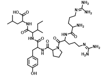
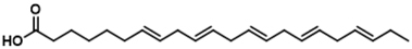
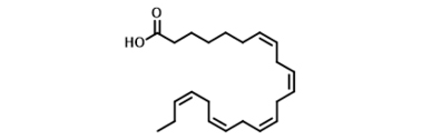
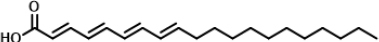
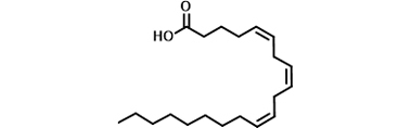
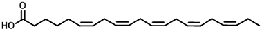
Two-dimensional structures of Neurotensin 8–13 and compounds from omega-3 fatty acids.
| No. | Structure | IUPAC name |
|---|---|---|
| 1 |
|
Neurotensin 8–13 |
| 2 |
|
alpha-linolenic acid |
| 3 |

|
Docosahexaenoic acid |
| 4 |
|
Docosapentaenoic acid |
| 5 |

|
Eicosapentaenoic acid |
| 6 |
|
Eicosatetraenoic acid |
| 7 |
|
Eicosatrienoic acid |
| 8 |
|
heneicosapentaenoic acid |
| 9 |

|
hexadecatrienoic acid |
| 10 |

|
stearidonic acid |
Preparation of ligand structure
Test ligand structures of the 9 compounds from omega-3 fatty acids and natural ligand are shown in Table
Preparation of protein receptor
The crystal structure of the neurotensin receptor crystal structure (PDB code 4GRV) was obtained from the PDB database (http/www/pdbbeta.rscb.org/pdb) with 2.80 Ǻ resolution. Neurotensin 8–13 is a natural ligand of the neurotensin receptor. The crystal structure of the neurotensin receptor used is neurotensin 8–13 and other residues. Neurotensin is a 13 amino acid neuropeptide that plays a role in the regulation of lutein hormone and prolactin release and also has interactions with the dopaminergic system. Plasma neurotensin levels in the intestine increase after fat digestion, we used neurotensin positions 8–13 in the neurotensin receptor crystal structure as posi- tions for all docking analyses.
Validation of the molecular docking method
Validation of the molecular docking method is performed by redocking a neurotensin 8–13 to a target protein that has been removed from the neurotensin receptor using the AutoDock Tools 1.5.6 program. The method is deemed successful if the root-mean-square deviation (RMSD) value returned is < 2 Å (
Docking simulation of the Neurotensin 8–13 and Test Ligands (compounds from omega-3 fatty acids)
The 3D structure of the Neurotensin 8–13 and Test Ligands were created and optimized using Chem3D Ultra 8.0. Structural optimization was carried out on the 3D structure of the reference and test ligands using the MM2 semi-empirical computational method. Calculations were carried out with geometry optimization on the minimum energy of the 3D structure of the compound to be used.
The structure of the neurotensin receptor (4GRV), neurotensin 8–13 and test ligands in the pdb format were converted into pdbqt format using the AutoDock Tools 1.5.6 program. The docking method was performed by tethering each ligand to neurotensin receptors using the tether coordinates (Grid Center) x = 40, y = 40, z = 40 Å and the Grid Box size coordinates x = 90.024, y = -11.196, and z = 68.538 Å. Each ligand was in a stable condition and interacted with biomacromolecules in a rigid condition.
Docking results were assessed for binding energy and chemical interactions such as hydrogen bonds, hydrophobic interactions and bond distances. These were visualized using the Discovery Studio Visualizer program. Discovery Studio is a comprehensive software suite for analyzing and modeling molecular structures, sequences, and other data of relevance to life science researchers. The product includes functionality for viewing and editing data along with tools for performing basic data analysis.
Molecular dynamic (MD) simulation
MD simulations to determine the ligand with the lowest binding energy at the neurotensin receptor (
Results and discussion
Preparation of protein receptor
The neurotensin receptor binds to natural ligands with chemical bonds. Natural ligands that interact with the neurotensin receptors (4GRV), namely neurotensin 8–13, were separated using Discovery Studio Visualizer software. The structure of the neurotensin receptor and neurotensin 8–13 are depicted in Fig.

Besides the Neurotensin receptor, exploration of the docking process requires a ligand. Ligand selection used in the process of tethering the target protein is based on initial screening results according to Lipinski’s Rule of Five. Ligands that are considered to have binding potential can enter the cell membrane to be absorbed by the body if they meet the following criteria: (1) molecular weight < 500 g/mol; (2) < 5 proton donor groups for hydrogen bonds; (3) < 10 proton acceptor groups for hydrogen bonds; and (4) a logarithmic value of the partition coefficient in water and 1-octanol < 5 (
Validation of molecular docking method
Analysis of the bonds formed between neurotensin 8–13 and the neurotensin receptor (4GRV) was performed using Discovery Studio Visualizer software. The analysis results of the bonds formed are shown in Table
| Protein | Compound | Binding energy (kcal/mol) | RMSD | Hydrogen bond distance (Ǻ) | Amino acids that bind | Nearest residues |
|---|---|---|---|---|---|---|
| 4GRV | Neurotensin 8–13 | -6.41 | 0.31 | 2.14; 2.36; 2.16 | TYR146; ARG213; PHE344 | VAL224 |
The molecular docking method is validated by redocking neurotensin 8–13 to the target protein. In this study, we redocked the neurotensin 8–13 to the neurotensin receptor (4GRV) at a resolution of 2.80 Ǻ. The redocking results had an RMSD value of 0.31 Å and a bond energy of -6.41 kcal/mol. According to (

Docking simulation of the Neurotensin 8–13 and Test Ligands (compounds from omega-3 fatty acids
Docking simulation of neurotensin 8–13 and Test Ligands (compounds from omega-3 fatty acids) was performed with the AutoDock Tools 1.5.6 program. The same coordinate settings at the site of the interaction between Neurotensin 8–13 and the neurotensin receptor (4GRV) were used for the docking simulation. This analysis was performed for binding energy. There was a hydrogen bond between the test/reference ligand and the neurotensin receptor. Docking simulation results are shown in Table
| Compound | Binding energy (kcal/mol) | Hydrogen bond distance (Ǻ) | Hydrogen bonds | Nearest amino acid residue(s) |
|---|---|---|---|---|
| alpha-linolenic acid | -7.0 | 1.91; 2.89; 1.99; 1.91 | ARG149; TYR251; ARG327; ARG328 | VAL224 |
| Docosahexaenoic acid | -8.96 | 2.15; 2.15; 2.18; 2.30; 2.00 | TYR351; ARG149; TYR146; ARG327; ARG328 | PHE344; TRY347; HIS348; TRP339 |
| Docosapentaenoic acid | -7.08 | 1.69; 2.08 | ARG327; ARG328 | PHE128; PHE344; TRY347; TRP339 |
| Eicosapentaenoic acid | -7.41 | 1.71; 2.03; 2.56 | ARG327; ARG149; TYR351 | HIS132; HIS348 |
| Eicosatetraenoic acid | -7.63 | 1.95; 1.78 | ARG149; ARG328 | PHE128; HIS348 |
| Eicosatrienoic acid | -7.24 | 2.55; 1.96; 1.96 | ARG327; TYR146; TYR351 | - |
| heneicosapentaenoic acid | -7.69 | 2.03; 2.06 | ARG327; ARG328 | TRP339 |
| hexadecatrienoic acid | -7.18 | 1.99; 2.01 | ARG327; ARG328 | TRP339 |
| stearidonic acid | -7.17 | 1.96; 2.09 | TYR351; ARG149 | PHE331; TRP339 |
Visualization of the docking interactions that occur between docosahexaenoic acid and receptors (4GRV) to the neurotensin receptor (4GRV) are shown in Fig.

Docking is a process of tethering interactions between ligands and proteins; it allows one to predict the position and orientation of ligands when bound to protein receptors (
Molecular dynamic simulation
The MD simulation was carried out using the chemical docosahexaenoic acid, eicosapentaenoic acid and heneicosapentaenoic acid, where the two test compounds had the same ΔG value. The RMSD and RMSF analysis of the receptor–ligand complex using GROMACS 2016 was carried out by measuring the stability of the RMSD and RMSF values in the system during the simulation (Fig.

RMSD analysis was used to assess the stability of the complex over time, while RMSF analysis assessed the stability per amino acid.
Assessed the stability per amino acid. Docosahexaenoic acid, eicosapentaenoic acid and heneicosapentaenoic acid, with the best docking score of the metabolites, was simulated with MD and its complex stability which are neurotensin receptor blockers. Docosahexaenoic acid, eicosapentaenoic acid and heneicosapentaenoic acid in complex with the neurotensin receptor showed the same high fluctuations. Meanwhile, the average RMSD fluctuations for each system, namely Docosahexaenoic acid, eicosapentaenoic acid, and heneicosapentaenoic acid, were 0.672, 0.437 and 0.650, respectively. The average RMSD showed that (eicosapentaenoic acid) had the lowest fluctuation, which indicates that the ligand has reached a stable conformation that binds to the protein (
SASA was analyzed for 100 ns of simulated MD trajectory, as shown in Fig.

To forecast the conformational changes in proteins that make them accessible to water molecules, the SASA was the result during simulations. Fig.
SASA was analyzed for 100 ns of simulated MD trajectory, as shown in Fig.
Polar solvation energy has a positive value while van der Waals, electrostatic, and SASA energies have a negative value in both of these complex systems. The results show that van der Waals, electrostatic, and SASA energy favor the binding while polar solvation energies oppose it in both complex systems. The total binding free energy of the ligands had varying values. Heneicosapentaenoic acid provided the lowest binding free energy -120.065 kJ/mol, while those for Docosahexaenoic acid, and eicosapentaenoic acid were -119.530, and -93.826 kJ/mol, respectively. The MM-PBSA analysis indicated that scopolin has better affinity for the neurotensin receptor. The binding free energy of the MD trajectories of the system complex was calculated using the MM-PBSA method for a timestep of 0–100 ns (Table
MM-PBSA energy summary ligand–neurotensin receptor during 100 ns simulation.
| Ligand | van der Waals energy (KJ/mol) | Electrostatic energy (KJ/mol) | Polar solvation energy (KJ/mol) | SASA energy (KJ/mol) | Total binding energy (KJ/mol) |
|---|---|---|---|---|---|
| Docosahexaenoic acid | -197.15 +/- 16.24 | -46.94 +/- 9.48 | 148.64 +/-15.46 | -24.075 +/- 1.27 | -119.53+/-16.20 |
| Eicosapentaenoic acid | -169.71 +/- 17.07 | -36.34 +/- 23.89 | 132.306 +/-30.29 | -20.088 +/- 1.43 | -93.83+/-16.61 |
| Heneicosapentaenoic acid | -184.44 +/- 15.28 | -46.94 +/- 11.49 | 131.710 +/- 15.27 | -20.388 +/- 0.94 | -120.07 +/-14.41 |
Conclusion
Based on the docking results, the ΔG values for omega-3 compounds of docosahexaenoic acid, eicosapentaenoic acid, heneicosapentaenoic acid and neurotensin 8–13 as native ligand were -8.96 ; -7.41 ; -6.34 and -6.41kcal/mol, respectively. Neurotensin 8–13 forms hydrogen bonds with TYR146, ARG213, and PHE344 of the neurotensin receptor, whereas docosahexaenoic acid forms hydrogen bonds with TYR146. Meanwhile, the average RMSD fluctuations for each system, namely docosahexaenoic acid, eicosapentaenoic acid, and heneicosapentaenoic acid, were 0.672, 0.437, and 0.650, respectively. The SASA of the neurotensin receptor-ligand complex on the graph for docosahexaenoic acid, eicosapentaenoic acid, and heneicosapentaenoic acid showed similar fluctuations. The average values for docosahexaenoic acid, eicosapentaenoic acid, and heneicosapentaenoic acid were 230.40, 229.89, and 230.20 nm2, respectively. This analysis correlated with that of the RMSD value, which indicated that eicosapentaenoic acid had better stability at the neurotensin receptor.
Acknowledgement
This research was supported by the Directorate of Research and Community Service of Universitas Padjadjaran through RDPD Grant 2023.
References
- Abdurrahman S, Ruslin R, Hasanah A, Mustarichie R (2021) Molecular docking studies and ADME-tox prediction of phytocompounds from Merremia peltata as a potential anti-alopecia treatment. Journal of Advanced Pharmaceutical Technology and Research 12(2): 132–39. https://doi.org/10.4103/japtr.JAPTR_222_20
- Abdurrahman S, Ruslin R, Hasanah AN, Mustarichie R, Ifaya M (2022) Active antialopecia Chemical identification of Merremia peltata leaves and computational study toward androgen receptor using molecular docking and molecular dynamic simulation. Scientific World Journal 2022: е1123047. https://doi.org/10.1155/2022/1123047
- Abdurrahman S, Ruslin R, Hasanah AN, Ifaya M, Mustarichie R (2023) Anti-alopecia activity of coumarin derivatives isolated from Merremia peltata leaves and computational study of their binding to androgen receptors using molecular docking and molecular dynamic simulation. Pharmaceuticals 16(5): 1–20. https://doi.org/10.3390/ph16050669
- Bowers LD (1989) High-performance liquid chromatography/mass spectrometry: State of the art for the drug analysis laboratory. Clinical Chemistry 35(7): 1282–1287. https://doi.org/10.1093/clinchem/35.7.1288
- Calder PC (2018) Very long-chain n-3 fatty acids and human health: Fact, fiction and the future. Proceedings of the Nutrition Society 77(1): 52–72. https://doi.org/10.1017/S0029665117003950
- Essmann U, Perera L, Berkowitz ML, Darden T, Lee H, Pedersen LG (1995) A smooth particle mesh Ewald method. The Journal of Chemical Physics 103(19): 8577–8593. https://doi.org/10.1063/1.470117
- Halder S, Kumar S, Sharma R (2013) The therapeutic potential of GPR120: A patent review. Expert Opinion on Therapeutic Patents 23(12): 1581–1590. https://doi.org/10.1517/13543776.2013.842977
- Huang F, del-Río-Navarro BE, Leija-Martinez J, Torres-Alcantara S, Ruiz-Bedolla E, Hernández-Cadena L, Barraza-Villarreal A, Romero-Nava R, Sanchéz-Muñoz F, Villafaña S, Marchat LA, Hong E (2019) Effect of omega-3 fatty acids supplementation combined with lifestyle intervention on adipokines and biomarkers of endothelial dysfunction in obese adolescents with hypertriglyceridemia. Journal of Nutritional Biochemistry 64: 162–169. https://doi.org/10.1016/j.jnutbio.2018.10.012
- Huei CS, Azlan A, Ismail A, Shafie NH, Sultana S (2020) Antioxidant and anti-obesity properties of local chilies varieties in Malaysia. Journal of Food Science and Technology 57(10): 3677–3687. https://doi.org/10.1007/s13197-020-04400-x
- Lipinski CA, Lombardo F, Dominy BW, Feeney PJ (2012) Experimental and computational approaches to estimate solubility and permeability in drug discovery and development settings. Advanced Drug Delivery Reviews 64(Suppl.): 4–17. https://doi.org/10.1016/j.addr.2012.09.019
- Mark P, Nilsson L (2001) Structure and dynamics of the TIP3P, SPC, and SPC/E water models at 298 K. Journal of Physical Chemistry A 105(43): 9954–9960. https://doi.org/10.1021/jp003020w
- Megawati G, Herawati D, Musfiroh I (2021) Binding affinity of omega 3 fatty acid as an agonist PPAR-γ and GPR120 receptor for obesity using molecular docking and ADME prediction. European Journal of Molecular & Clinical Medicine 7(10): 1686–1695.
- Mohamed GA, Ibrahim SRM, Elkhayat ES, El Dine RS (2014) Natural anti-obesity agents. Bulletin of Faculty of Pharmacy, Cairo University 52(2): 269–284. https://doi.org/10.1016/j.bfopcu.2014.05.001
- Morgan PJ, Young MD, Lloyd AB, Wang ML, Eather N, Miller A, Murtagh EM, Barnes AT, Pagoto SL (2017) Involvement of fathers in pediatric obesity treatment and prevention trials: A systematic review. Pediatrics 139(2): e20162635. https://doi.org/10.1542/peds.2016-2635
- Musfiroh I, Megawati G, Herawati DMD, Ifaya M (2022) Stability of omega-3 compounds complex with Ppar-γ receptor as an anti-obesity using molecular dynamic simulation. International Journal of Applied Pharmaceutics 14(Special Issue 5): 45–49. https://doi.org/10.22159/ijap.2022.v14s5.04
- Musfiroh I, Megawati G, Herawati DMD, Rusdin A (2021) 3D-pharmacophore modelling of omega-3 derivatives with peroxisome proliferator-activated receptor gamma as an anti-obesity agent. International Journal of Applied Pharmaceutics 13(Special Issue 4): 167–170. https://doi.org/10.22159/ijap.2021.v13s4.43851
- Psimadas D, Georgoulias P, Valotassiou V, Loudos G (2012) Molecular nanomedicine towards cancer. Journal of Pharmaceutical Sciences 101(7): 2271–2280. https://doi.org/10.1002/jps.23146
- Ramírez D, Caballero J (2018) Is it reliable to take the molecular docking top scoring position as the best solution without considering available structural data? Molecules 23(5): 1–17. https://doi.org/10.3390/molecules23051038
- Salam Md M, Yousuf R, Salam Md W, Haque M (2023) Obesity and overweight: A global public health issue. Advances in Human Biology 13(1): 154–156.
- Song T, Yang Y, Zhou Y, Wei H, Peng J (2017) GPR120: A critical role in adipogenesis, inflammation, and energy metabolism in adipose tissue. Cellular and Molecular Life Sciences 74(15): 2723–2733. https://doi.org/10.1007/s00018-017-2492-2
- Sousa Da Silva AW, Vranken WF (2012) ACPYPE – AnteChamber PYthon parser interface. BMC Research Notes 5: 1–8. https://doi.org/10.1186/1756-0500-5-367
- Veber DF, Johnson SR, Cheng HY, Smith BR, Ward KW, Kopple KD (2002) Molecular properties that influence the oral bioavailability of drug candidates. Journal of Medicinal Chemistry 45(12): 2615–2623. https://doi.org/10.1021/jm020017n