Research Article |
|
Corresponding author: Reem A. Issa ( r.issa@ammanu.edu.jo ) Corresponding author: Mahmoud Abu-Samak ( m_abusamak@asu.edu.jo ) Academic editor: Rumiana Simeonova
© 2024 Raheeq Khairy Abdullah, Reem A. Issa, Mahmoud Abu-Samak, Beisan A. Mohammad, Manal A. Abbas, Shady Helmi Awwad.
This is an open access article distributed under the terms of the Creative Commons Attribution License (CC BY 4.0), which permits unrestricted use, distribution, and reproduction in any medium, provided the original author and source are credited.
Citation:
Abdullah RK, Issa RA, Abu-Samak M, Mohammad BA, Abbas MA, Awwad SH (2024) Nephroprotective effects of Equisetum ramosissimum L. extract in streptozotocin-induced diabetic rats. Pharmacia 71: 1-11. https://doi.org/10.3897/pharmacia.71.e113659
|
Abstract
Diabetes is a widespread health issue that impacts people all over the globe. The Equisetum ramosissimum L. plant has numerous traditional uses and pharmacological properties, including antidiabetic effects.The objective of this study was to thoroughly examine the advantages of incorporating extracts from the aerial components of E. ramosissimum to control diabetic nephropathy. The phytochemical constituents of E. ramosissimum extract were explored using phytochemical and HPLC analysis, focusing on phenols and flavonoid content. The effect of plant extract was evaluated on different kidney function parameters linked to diabetic nephropathy (fasting blood glucose, creatinine, uric acid, and urea) in streptozotocin induced-diabetic rats. Histopathological changes in the liver were also examined. The results showed methanol and ethanol extracts of E. ramosissimum have a total content of phenols (equivalent to gallic acid, 7.62 and 8.97 mg/g) and flavonoids (equivalent to quercetin, 8.87 and 12.86 mg/g), respectively. After conducting the UHPLC-MS/MS analysis, it was found that both the methanol and ethanol extracts contained isoferulic acid, ISO-Orientin, myristic acid, linoelaidic acid, rutin, and 3-Glu-7-Rha quercetin. Additionally, isoferulic acid, myristic acid, linoelaidic acid, rutin, and 3-Glu-7-Rha quercetin were present in the extracts. The ethanol extract of E. ramosissimum significantly impacted STZ-induced diabetic mice by reducing their fasting blood glucose levels, and their creatinine and urea levels (P < 0.005). In conclusion, E. ramosissimum ethanol extract has shown potential effects to counteract some diabetes consequences on kidney function. Therefore, further studies are required to investigate its effect on other diabetes-related complications.
Keywords
Equisetum ramosissimum, diabetes, nephroprotective, kidney functions, phenols, flavonoids
Introduction
According to the WHO report (
The persistent complications of diabetes are broadly partitioned into microvascular and macrovascular complications, including diabetic nephropathy, which influences 20–30% of diabetic patients, with oxidative harm playing a critical part in its pathogenesis (Al-Waili et al. 2017).
Herbal and dietary supplements that are rich in flavonoids can potentially decrease the occurrence of diabetes. This effect is achieved by lowering blood sugar levels and offering other benefits that aid in the treatment and prevention of complications related to diabetes (
Some flavonoid compounds have shown preventive impacts on diabetic complications, such as hesperidin, naringenin, quercetin kaempferol, puerarin, and myricetin (Gandhi et al. 2020). The therapeutic effects of numerous components of flavonoids on diabetic nephropathy have been illustrated in vitro and in vivo studies, particularly apigenin, baicalein, and catechin, through anti-oxidation effects (
The study species Equisetum ramosissimum is called “horsetail” (
Extracts from various Equisetum species were utilized in recent studies on diabetic rats. The results indicate that E. arvense exhibits high levels of phenolic and flavonoid compounds with potent antioxidant properties, making it a promising addition to diabetes therapy (
The main goal of this research was to explore the potential health benefits of different extract preparations from E. ramosissimum to prevent diabetes-related nephropathy. Multiple extracts of E. ramosissimum were created and analyzed to determine their antioxidant properties, concentration of phenols, and flavonoid content. The plant extract with optimal content of phytocomponents was investigated for its effect on different kidney function parameters linked to diabetes nephropathy, as it is considered a major complication of diabetes.
Materials and methods
Preparation of plant materials
For this study, the E. ramosissimum species was sourced from the Mujib reserve in Jordan. The plant species was authenticated by a knowledgeable botanist from the Royal Society for the Conservation of Nature (RSCN) with voucher number (E. ramosissimum 5/7/2017). The aerial components of the E. ramosissimum were cleaned, dried, and ground into smaller pieces using a blender.
The extraction method
A range of extracts from the plant under study was obtained using the maceration method. The extracts were created by mixing 500 mL of either methanol or ethanol with 50 g of dry plant material at room temperature (25.0 °C) for 24 hours. Then, the resulting mixture was filtered with filter paper. Afterwards, the extracts were completely dehydrated using a rotary evaporator (R-300, Buchi, USA), at 60 °C and 90 rpm. Finally, each dried extract was accurately weighed to determine the extraction yield.
Determination of total phenolic content
The total phenol content was determined through the Folin-Ciocalteu technique, as outlined in Sadeghi et al.’s publication (2015). A combination of 5 mL of plant extract solution (or gallic acid), Folin-Ciocalteu reagent, and 5% aqueous Na2CO3 was prepared. After a 30-minute incubation period, the phenolic concentration was measured calorimetrically using Spectrophotometer (Hitachi U-1800, UK) at 765 nm. The total phenolics (mg/mL) amount was subsequently calculated as the gallic acid equivalent.
Identification of phenols and flavonoids using UHPLC/MS-MS
After dissolving each extracted sample in 2 mL of DMSO and up to 50 ml of acetonitrile, they were centrifuged at 4000 rpm for 2.0 min. Then, 3.0 µL of each sample containing 1.0 ml was injected into the autosampler.
A Bruker Daltonik (Bremen, Germany) Impact II electrospray ionization quadrupole time of flight (ESI-Q-TOF) system equipped with a Bruker Daltonik Elute Ultra-Performance Liquid Chromatography (UPLC) system (Bremen, Germany) were thoroughly utilized to test specific compounds against an integrated library. Chromatography was employed to separate the compounds, and they were analyzed for m/z and the retention time using highly precise Bruker TOF MS and standards.
Chromatographic separation was performed with the utmost precision using the Bruker solo 2.0 C-18 UHPLC column (100 mm × 2.1 mm × 2.0 m) at a consistently reliable flow rate of 0.51 mL/min and a stable column temperature of 40 °C. The solvents used, a mixture of water containing 0.05% formic acid (A) and acetonitrile (B), were carefully selected to optimize the results. B’s method was skillfully employed for a 35-minute analysis with gradient elution (80–95%), ensuring the accuracy and efficiency of the process.
The experimental study design
Animals
Wistar rats weighing 150–200 grams and aged 70 days received a regular diet and had unrestricted water access from Hammoudeh business in Amman, Jordan. The experiments on rats were conducted in accordance with the Declaration of Helsinki. Before the experiment, the rats were acclimated to the animal housing conditions for ten days, including a temperature range of 21–23 °C and a 12-hour light-dark cycle. The Institutional Committee on Ethics of Animal Experimentation approved the procedure. All procedures were performed in accordance with international regulations for the care and use of laboratory animals. Ethical approval for the study was obtained by the Institutional Review Board at Applied Science Private University, Amman, Jordan, Approval Number: 2021-PHA-40.
Induction of diabetes in rats
The rats were given two doses of freshly dissolved streptozotocin (STZ, Sigma, USA). through intraperitoneal (IP) administration. A total of 55 mg/kg of STZ was given in citrate buffer (0.01 M, pH 4). The first dose was 30 mg/kg, followed by a second dose of 25 mg/kg on the third day after 48 hours. After each STZ dose, the rats were returned to their cages and provided unrestricted food and drink access (5% dextrose to prevent severe hypoglycemia). Control rats were given citrate buffer.
After 72 hours of administering STZ, the rats were confirmed to have diabetes (STZ-DM: STZ-induced diabetes). A commercial glucose kit measured their blood glucose levels Qualigens Diagnostics – AccuChek- (ROCHE, TD-4277, Augsburg, Germany). The test of the blood sample from the rat’s tail was applied twice on each test-strip lot. If the fasting blood glucose (FBG) level was 250 mg/dL or higher (
Two more samples were collected and analyzed alongside the blood droplets to confirm and monitor STZ-DM as follows:
- Three days after the STZ injection and before the therapy, the blood samples were collected from the rats. The retro-orbital plexus was accessed to collect 0.2 mL from each animal into heparinized tubes. The collected plasma was centrifuged for 10 minutes at 2000 rpm and tested for FBG.
- The animals had blood samples taken at the end of the 21-day experiment. Each sample had a 2–3 ml volume and was stored in tubes with heparin. The tubes were allowed to coagulate for an hour and then centrifuged at 4000 rpm for 10 minutes. At least 1 ml of blood was transferred into Eppendorf tubes to extract serum, labelled accordingly. All samples remained stored at -80 °C until they were ready for biochemical testing.
Plant extract solution preparation and administration
The dried plant extract was dissolved in distilled water at 100 or 150 mg/ml concentrations to create fresh ethanol E. ramosissimum extract stock solutions. Each was given orally to rats through an intragastric tube, with a dose of 200 or 300 mg/kg BW, like a prior research study (Carneiro 2013). The E. ramosissimum extract treatments began on the third day after STZ injection, and the experiment was conducted for three weeks on the same days after diabetes was induced.
Animal groups
A detailed investigation examined the potential benefits of E. ramosissimum extract in preventing diabetes and protecting against kidney damage. The study involved forty-two rats, divided into seven groups of six animals, each as follows:
- Group (C): non-diabetic control group.
- Group (SDM): STZ-induced diabetic rats (55 mg/kg).
- Group (SDMET): STZ-induced diabetic rats, orally treated with the standard antidiabetic drug metformin (50 mg/kg/day).
- Group (Q2): Non-diabetic rats received E. ramosissimum ethanol extract (200 mg/kg/day).
- Group (SDMQ2): STZ-induced diabetic rats received E. ramosissimum ethanol extract (200 mg/kg/day).
- Group (Q3): Non-diabetic rats received E. ramosissimum ethanol extract (300 mg/kg/day).
- Group (SDMQ3): STZ-induced diabetic rats received E. ramosissimum ethanol extract (300 mg/kg/day).
Biochemical analysis
The rats were not fed the night before they were euthanized on the 21st day of the experiment. Blood samples were collected before and after the experiment to produce serum, which was then analyzed using liquicolor-HUMAN kits to measure the levels of FBG, creatinine (Cr), uric acid (UA), and urea (U).
Histopathological studies
The kidney slices, fixed in formalin and embedded in paraffin wax, were stained using Hematoxylin and Eosin (H & E) at a thickness of 4 micrometers. The sections were thoroughly examined under a Leica microscope (The Life Sciences companies at Danaher Corporation (“Danaher”), Germany) and captured using a Leica MC 170 HD camera utilizing LAS EZ software.
Statistical analysis
Version 27.0 of the Statistical Package for the Social Sciences (SPSS) for Windows, developed in Chicago, IL, USA, was utilized to perform the statistical analysis.
The one-way ANOVA test was used to identify any significant differences in the average values of each parameter among different experimental groups. The post hoc multiple comparisons with Dunnett’s Multiple Comparisons tests were used to analyze any meaningful variations in mean values between the experimental and control groups.
The study aimed to thoroughly examine the potential impact of FBG as a predictor of diabetes mellitus and Cr, U, and UA as predictors of diabetes nephropathy from the beginning to the end of the trial. Using a stepwise approach to multiple linear regression, researchers determined the factors that played a crucial role in the relationships between diabetes and diabetic nephropathy across various research groups.
Results
Plant sample extraction
The extraction yield was computed using % w/w dry weight. Based on the data, it was found that ethanol produced the highest output at 3.42%, followed closely by methanol at 2.78%.
Determination of total phenolic content
Based on the analysis of total phenol content, it has been determined that the methanol and ethanol extracts contain 7.62 and 8.97 mg/g, equivalent to gallic acid, respectively.
Determination of total flavonoid content
Upon evaluating their total flavonoid content, it has been determined that the methanol and ethanol extracts possess 8.87 and 12.86 mg/g of quercetin, respectively.
Identification of phenols and flavonoids using UHPLC/MS-MS
Identification of the phytocomponents present in the methanol and ethanol extracts of E. ramosissimum was made possible using UHPLC/MS-MS. Tables
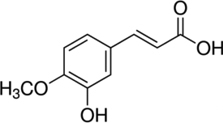
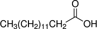
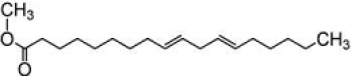
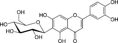
Phytochemicals detected in E. ramosissimum methanol extract using UHPLC/MS-MS.
| Compound name | Structure | RT [min] | m/z |
|---|---|---|---|
| 3-Hydroxy-4-methoxycinnamic acid (Iso ferulic acid) |
|
5.39 | 173.16261 |
| Myristic acid |
|
27.14 | 181.18303 |
| Linoelaidic acid |
|
29.82 | 213.26471 |
| ISO-Orientin |
|
4.88 | 257.37702 |
| Rutin |
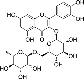
|
5.6 | 277.42807 |
| 3-Glu-7-Rha Quercetin |
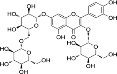
|
5.25 | 281.43828 |
Phytochemicals detected in E. ramosissimum ethanol extract using UHPLC/ MS-MS.
| Compound name | Structure | RT [min] | m/z |
|---|---|---|---|
| 7-Glu Chrysoeriol |
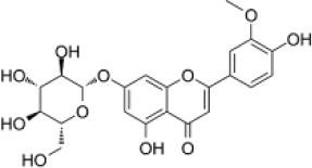
|
7.1 | 261.38723 |
| 7-Methoxy-4’-hydroxyflavone |
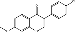
|
11.43 | 193.21366 |
| Succinic acid |
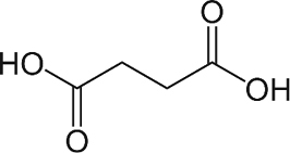
|
0.98 | 117.01967 |


Biochemical changes within study groups at baseline and the end of the study period
Changes in body weight
During the study, all groups, except the control group, showed a considerable reduction in their average body weight at the beginning and end of the research. This fact is undeniably supported by the P-value of 0.05, as shown in Table
Changes of BW at baseline and the end of experiment in all study groups.
| Body weight (g) | ||||
|---|---|---|---|---|
| Group (n=6) | Baseline | End of the study | Change (%) | P a |
| C | 178.3±12.2 | 190.8±10.7 | 12.50 (7.01%) | < 0.001 |
| SDM | 210±22.6 | 142.5±24.7 | -67.50 (-32.14%) | < 0.001 |
| SDMET | 200±16.1 | 166.7±8.2 | -33.33 (-16.67%) | 0.06 |
| Q2 | 205±5.5 | 192.5±5.2 | -12.5 (-6.01%) | < 0.001 |
| SDMQ2 | 221.7±15.1 | 152.5±32.1 | -69.17 (-31.20%) | 0.002 |
| Q3 | 187.5±10.4 | 175±10.5 | -12.5 (-6.67%) | < 0.001 |
| SDMQ3 | 213.3±15.4 | 167.5±26.2 | -45.83 (-21.5%) | 0.001 |
| F | 6.367 | 5.419 | ||
| P b | < 0.001 | |||
| P c | < 0.001 | |||
Changes in fasting blood glucose
Based on the results shown in Table
Changes of FBG at baseline and the end of the experiment in all study groups.
| Fasting Blood Glucose (FBG) (mg/dl) | ||||
|---|---|---|---|---|
| Group (n=6) | Baseline | End of the study | Change (%) | P a |
| C | 93.90±5.04 | 93.78±3.84 | -0.06 (-0.06%) | 0.955 |
| SDM | 487.8±40.4 | 501.7±64.6 | 43.83 (8.99%) | 0.104 |
| SDMET | 326±85.8 | 81.2±27.4 | -244.78 (-75.08%) | < 0.001 |
| Q2 | 91±8.2 | 90.9±9.8 | -0.13 (-0.14%) | 0.979 |
| SDMQ2 | 423±115.7 | 132.7±55.6 | -290.3 (-68.7) | |
| Q3 | 93.4±14.5 | 101.1±8.2 | 7.72 (8.27%) | 0.369 |
| SDMQ3 | 259.2±74.7 | 155.93±78.1 | -103.27 (-39.8) | 0.002 |
| F | 37.93 | 66.714 | ||
| P b | < 0.001 | |||
| P c | < 0.001 | |||
Changes in urea levels
As shown in Table
Changes in urea (U) at baseline and the end of the experiment in all study groups.
| Urea (U) (ng/ml) | ||||
|---|---|---|---|---|
| Group (n=6) | Baseline | Follow-up | Change (%) | P a |
| C | 37.7±4.04 | 37.5±5.14 | -0.2 (-0.53%) | 0.91 |
| SDM | 42.6±5.2 | 43.1±0.2 | 0.5 (1.17%) | 0.71 |
| SDMET | 43±9.3 | 53.1±9.5 | 10.02 (23.3%) | 0.012 |
| Q2 | 40.3±2.6 | 34±4 | -6.23 (-15.46%) | 0.007 |
| SDMQ2 | 43±9.34 | 32.5±4.9 | -10.6 (-24.65%) | 0.013 |
| Q3 | 39.8±1.9 | 29.9±3.4 | -9.93 (-24.25%) | 0.003 |
| SDMQ3 | 30.6±5.2 | 30.6±11 | 0.00 (0%) | 0.995 |
| F | 4.6 | 9.2 | ||
| P b | 0.002 | |||
| P c | < 0.001 | |||
Fig.

Final Mean values of Urea serum levels at the end of the study. Abbreviations: C = non-diabetic control group, SDM = STZ-induced diabetic rats, SDMET = STZ-induced diabetic rat + metformin (50 mg/kg/day), Q2 = Low dose extract (200 mg/kg/day), SDMQ2 = STZ-induced diabetic rats + Low dose extract (200 mg/kg/day), Q3 =: High dose extract (300 mg/kg/day), SDMQ3 = STZ-induced diabetic rats + High dose extract (300 mg/kg/day). NOTE: Results are represented as Mean±SD (n=6 for each group). * p < 0.05, ** p < 0.01, and *** p < 0.001 when compared with the SDM; • p < 0.05, •• p < 0.01, and •••p < 0.001 when compared with the SDMT (Dunnett’s Multiple Comparisons Test).
Changes in creatinine levels
As presented in Table
Changes in mean creatinine (Cr) levels at baseline and the end of the experiment in all study groups.
| Creatinine (Cr) (ng/ml) | ||||
|---|---|---|---|---|
| Group (n=6) | Baseline | Follow-up | Change (%) | P a |
| C | 0.44±0.05 | 0.43±0.08 | -0.08 (-18.18%) | 0.94 |
| SDM | 0.51±0.07 | 0.62±0.07 | 0.11 (21.57%) | 0.05 |
| SDMET | 0.44±0.05 | 0.55±0.05 | 0.11 (25%) | 0.035 |
| Q2 | 0.43±0.04 | 0.50±0.02 | 0.08 (18.60%) | 0.004 |
| SDMQ2 | 0.62±0.07 | 0.48±0.06 | -0.13 (-20.97%) | 0.001 |
| Q3 | 0.44±0.02 | 0.43±0.04 | -0.02 (-4.54%) | 0.413 |
| SDMQ3 | 0.51±0.07 | 0.42±.0.12 | -0.09 (-17.65%) | 0.278 |
| F | 8.589 | 7.301 | ||
| P b | < 0.001 | |||
| P c | < 0.001 | |||

Final Mean values of Urea serum levels at the end of the study. Abbreviations: C = non-diabetic control group, SDM = STZ induced diabetic rats, SDMET = STZ-induced diabetic rat + metformin (50 mg/kg/day), Q2 = Low dose extract (200 mg/kg/day), SDMQ2 = STZ-induced diabetic rats + Low dose extract (200 mg/kg/day), Q3 =: High dose extract (300 mg/kg/day), SDMQ3 = STZ-induced diabetic rats+ High dose extract (300 mg/kg/day). NOTE: Results are represented as Mean±SD (n=6 for each group). * p < 0.05, ** p < 0.01, and *** p < 0.001 when compared with the SDM; • p < 0.05, •• p < 0.01, and •••p < 0.001 when compared with the SDMT (Dunnett’s Multiple Comparisons Test).
Changes in UA levels
Table
Changes in Uric acid (UA) at baseline and the end of the experiment in all study groups.
| Uric Acid (UA) (ng/ml) | ||||
|---|---|---|---|---|
| Group (n=6) | Baseline | Follow-up | Change (%) | P a |
| C | 1.17±0.13 | 1.34±0.18 | 0.17 (14.52%) | 0.64 |
| SDM | 1.67±0.11 | 1.86±0.37 | 0.19 (11.38%) | 0.352 |
| SDMET | 1.17±0.13 | 1.18±0.39 | 0.01 (0.85%) | 0.948 |
| Q2 | 1.11±0.09 | 1.51± 0.51 | 0.41 (36.94%) | 0.126 |
| SDMQ2 | 1.86±0.37 | 1.72±0.26 | -0.14 (-7.53%) | 0.234 |
| Q3 | 1.20±0.16 | 1.21±0.25 | 0.01 (0.003%) | 0.948 |
| SDMQ3 | 1.67±0.11 | 1.00±0.24 | -0.67 (-40.12%) | 0.003 |
| F | 17.980 | 5.858 | ||
| P b | < 0.001 | |||
| P c | < 0.001 | |||
Stepwise regression analysis
The findings from Table
The multivariate associations between the experiment variables using stepwise regression at the end of the experiment (3 weeks).
| Dependent variable | Study group | Univariate effect estimate | Coefficient | ||||
|---|---|---|---|---|---|---|---|
| B | F | R | R2 | P-value | |||
| Cr | SDMQ2 | FBG | 0.002 | 13.134 | 0.876 | 0.767 | 0.022 |
| BW | 0.001 | 26.723 | 0.973 | 0.947 | 0.012 | ||
| UA | SDMQ2 | BW | -0.007 | 7.901 | 0.815b | 0.664 | 0.048 |
| SDMQ3 | U | 0.02 | 21.964 | 0.920 | 0.846 | 0.009 | |
The stepwise regression analysis showed significant associations between IDVs such as FBG, BW, and Cr levels. In addition, the influence of BW on UA levels was also observed. During follow-up, the multiple linear regression model explained a considerable 95% and 66.4% of the variation in Cr and UA, respectively. The SDMQ3 showed a robust reciprocal association between U and UA levels, with a whopping 85% of the variation in UA or U levels at follow-up fully explained by the model.
Histopathological studies
The control group’s rat liver tissue samples underwent light microscopy analysis to reveal the typical histological architecture of the hepatocytes, central vein, sinusoids, hepatic triads, and Kupffer cells. However, diabetic rats’ liver tissue samples exhibited necrotic hepatocytes with lymphocyte infiltration, disorganized sinusoids, and dilated central veins. It is important to note that compared to metformin-treated rats, diabetic rats treated with the extract showed significantly reduced necrosis in the liver, as seen in Fig.

Under light microscopy, the kidney sections of control rats displayed regular Bowman’s capsule, proximal tubules, and distal tubules. However, STZ-treated diabetic rats showed renal abnormalities, including glomerular tuft atrophy, renal tubule degeneration, and an increase in Bowman’s gap size, as well as hemorrhage and renal blood vessel congestion. Despite most administered Bowman’s capsules to diabetic rats with typical kidney damage, some distal tubules exhibited widening. Regrettably, metformin did not provide any protection against STZ-induced kidney damage, as shown in Fig.

Kidney sections (A) control. B, C. STZ-treated diabetic-rat showing atrophy of glomerular tuft (white arrow in B arrow), increase in the Bowman’s space area and degeneration of renal tubules (Black arrow in C) associated with severe hemorrhage and congestion of renal blood vessels; D. Extract-treated rats. Renal tubules (Black arrow), Bowman’s capsule (white arrow); E, F. Metformin-treated rats. H & E stain.
Discussion
The Jordanian herbal treatments for diabetes were thoroughly examined by
According to preclinical studies, it is evident that E. ramosissimum ethanol extract (
This study has shown that E. ramosissimum extract has similar effects to metformin in reducing FBG, U and Cr levels in diabetic and normal rats. It is noteworthy that previous studies have identified several Equisetum plant species that protect against adverse impacts on renal function in diabetic rats (
In SDMQ2 group, significant reduction (P< 0.05) in the means serum U and Cr levels were observed, while means serum UA levels was reduced in SDMQ3 only. These findings may point to potential powerful antioxidant activity for the extracts, but higher doses as in SDMQ3 study group might have an adverse or toxic effects (
Furthermore, the same group (SDMQ2) practically showed similar effect to that observed in diabetic -metformin treated group (SDM). Accordingly, in the term of the potential anti-DN effects for E. ramosissimum extract observed in SDMQ2 group , its bioactive compounds are likely to have therapeutic outcomes . Based on the stepwise regression findings in these experiments, the bioactive compound in the extract is expected to act peripherally rather than centrally. in this context, it has shown that the intervention of E. myriochaetum extract significantly reduced FBG levels without significant changes in the insulin levels in type 2 diabetic patients (
The antidiabetic and nephron protective effect for E. ramosissimum observed in the present study can be explained by various phytochemicals, such as phenols and flavonoids, with pronounced antioxidant effects (
In addition, the HPLC analysis for the extract revealed the presence of ferulic acid isomers (isoferulic acid). This compound is well known to prevent the synthesis of advanced glycation end products, which is responsible for altering other proteins and generating oxidizing intermediates, causing oxidative stress in vascular cells and other tissues. As a result, this phytocomponent may play an essential preventive role in age-related illnesses and diabetic complications, including nephropathy (
The fatty acids, linolelaidic and myristic acids were observed in the prepared extracts. Compositions of fatty acids in four Equisetum sp. (E. arvense, E. palustre, E. ramosissimum, and E. telmateia) revealed that linoleic acid was the predominant fatty acid in these species. In contrast, myristic acid was the second major fatty acid (
Conclusions
Accordingly, the antidiabetic and nephron protective effects observed in STZ-induced diabetic rats treated with the ethanolic extract of E. ramosissimum can be considered a novel finding. A significant reducing effect was observed on FBG, Cr, and U levels in STZ-induced diabetic rats.. This expectation was raised from the significant association between BW and FBG with UA and Cr levels in the STZ-diabetic rats treated with different doses of the extract.
The high content of phenols and flavonoids in the ethanol extracts of E. ramosissimum is likely to be responsible for the proposed effect via antioxidant activity. Therefore, further research is required to investigate its mechanism of action on the molecular level.
Acknowledgements
The authors thank Mr Salim for his technical assistance and Mr Anas Sabarini for preparing the specimen and verifying the plant. The Applied Science Private University/Faculty of Pharmacy in Amman, Jordan, is acknowledged for providing the necessary infrastructure and research facilities. Furthermore, the Deanship of Graduate Studies at Al-Ahliyya Amman University in Amman, Jordan, has funded this work.
References
- Afifi FU, Kasabri V (2013) Pharmacological and phytochemical appraisal of selected medicinal plants from Jordan with claimed antidiabetic activities. Scientia Pharmaceutica 81(4): 889–932. https://doi.org/10.3797/scipharm.1212-20
- Al-Bayati M, Issa R, Abu-Samak M, Alnsour L, Awwad S (2023) Phytochemical analysis and evaluation of anti-hyper lipidaemic effect for ethanolic leaf extract of Equisetum ramosissimum L.: In vivo study on rats’ models. Pharmacia 70(3): 557–568. https://doi.org/10.3897/pharmacia.70.e101623
- Alkhalidy H, Wang Y, Liu D (2018) Dietary flavonoids in the prevention of T2D: An overview. Nutrients 10(4): e438. https://doi.org/10.3390/nu10040438
- Al-Matubsi HY, Oriquat GA, Abu-Samak M, Al Hanbali OA, Salim MD (2016) Effects of lipoic acid supplementation on activities of cyclooxygenases and levels of prostaglandins E2 and F2α metabolites, in the offspring of rats with streptozotocin-induced diabetes. Journal of Diabetes Research 2016: e9354937. https://doi.org/10.1155/2016/9354937
- Alves CFDS, Bonez PC, de Souza MDE, da Cruz RC, Boligon AA, Piana M, Brum TF, Rossi GG, Jesus RDS, Grando TH, Monteiro SG, de Campos MMA, Giongo JL, Santos RCV (2016) Antimicrobial, antitrypanosomal and antibiofilm activity of Equisetum hyemale. Microbial Pathogenesis 101: 119–125. https://doi.org/10.1016/j.micpath.2016.11.008
- Ang L, Walkowiak R, Chan W (2015) “Short taxonomic notes on genus equisetum (horsetail)”, the international equisetological association and world equisetum program – research service. https://doi.org/10.13140/RG.2.2.29285.60644
- Angel GM, Gallardo-Hernández AG, Altamirano-Bustamante M, de la Chesnaye-Caraveo E (2020) Chronic hypoglycemic and hypolipidemic effect of Equisetum myriochaetum aerial parts on streptozotocin-induced diabetic rats. Pharmacology Online 2: 200–212.
- Bai L, Li X, He L, Zheng Y, Lu H, Li J, Zhong L, Tong R, Jiang Z, Shi J, Li J (2019) Antidiabetic potential of flavonoids from traditional Chinese medicine: A review. The American journal of Chinese medicine 47(05): 933–957. https://doi.org/10.1142/S0192415X19500496
- Banerjee SK, Mukherjee PK, Maulik SK (2003) Garlic as an antioxidant: the good, the bad and the ugly. Phytotherapy Research 17(2): 97–106. [PMID: 12601669] https://doi.org/10.1002/ptr.1281
- Barham A, Mohammad B, Hasoun L, Awwad S, Mosleh I, Aljaberi A, Abu-Samak M (2021) The combination of omega-3 fatty acids with high doses of vitamin D3 elevate A1c levels: A randomized clinical trial in people with vitamin D deficiency. International Journal of Clinical Practice 75(11): e14779. https://doi.org/10.1111/ijcp.14779
- Bhebhe M, Füller TN, Chipurura B, Muchuweti M (2016) Effect of solvent type on total phenolic content and free radical scavenging activity of black tea and herbal infusions. Food Analytical Methods 9(4): 1060–1067. https://doi.org/10.1007/s12161-015-0270-z
- Boeing T, Moreno KGT, Gasparotto Junior A, da Silva LM, de Souza P (2021) Phytochemistry and pharmacology of the genus Equisetum (Equisetaceae): A narrative review of the species with therapeutic potential for kidney diseases. Evidence-Based Complementary and Alternative Medicine 2021: e6658434. https://doi.org/10.1155/2021/6658434
- Carneiro DM, Tresvenzol LMF, Jardim PCBV, Cunha LCD (2013) Equisetum arvense: scientific evidences for clinical use. International Journal of Biology, Pharmacy, and Allied Sciences 2(8): 1579–1596.
- Cetto AA, Wiedenfeld H, Revilla MC, Sergio IA (2000) Hypoglycemic effect of Equisetum myriochaetum aerial parts on streptozotocin diabetic rats. Journal of Ethnopharmacology 72(1–2): 129–133. https://doi.org/10.1016/S0378-8741(00)00218-X
- Do QD, Angkawijaya AE, Tran-Nguyen PL, Huynh LH, Soetaredjo FE, Ismadji S, Ju Y-H (2014) Effect of extraction solvent on total phenol content, total flavonoid content, and antioxidant activity of Limnophila aromatica. Journal of Food and Drug Analysis 22(3): 296–302. https://doi.org/10.1016/j.jfda.2013.11.001
- Fons F, Froissard D, Bessière JM, Fruchier A, Buatois B, Rapior S (2013) Volatile composition of six horsetails: Prospects and perspectives. Natural Product Communications 8(4): 509–512. https://doi.org/10.1177/1934578X1300800426
- Gheith O, Farouk N, Nampoory N, Halim MA, Al-Otaibi T (2016) Diabetic kidney disease: world wide difference of prevalence and risk factors. Journal of Nephropharmacology 5(1): 1–49. https://doi.org/10.4103/1110-9165.197379
- Hegedűs C, Muresan M, Badale A, Bombicz M, Varga B, Szilágyi A, Sinka D, Bácskay I, Popoviciu M, Magyar I, Szarvas MM, Szőllősi E, Németh J, Szilvássy Z, Pallag A, Kiss R (2020) SIRT1 activation by Equisetum arvense L. (Horsetail) modulates insulin sensitivity in streptozotocin induced diabetic rats. Molecules 25(11): e2541. https://doi.org/10.3390/molecules25112541
- Hua C, Linling L, Feng X, Yan W, Honghui Y, Conghua W, Shaobing W, Zhiqin L, Juan H, Yuping W, Shuiyuan C, Fuliang C (2013) Expression patterns of an isoflavone reductase-like gene and its possible roles in secondary metabolism in Ginkgo biloba. Plant Cell Reports 32(5): 637–650. https://doi.org/10.1007/s00299-013-1397-2
- Hunt D, Hemmingsen B, Matzke A, Varghese C, Hammerich A, Luciani S, Hennis A, Branca F, Bull F, Berdzuli N, Dangou J-M, Pendse R, Pratt A, Mikkelsen B (2021) The WHO global diabetes compact: A new initiative to support people living with diabetes. The Lancet Diabetes & Endocrinology 9(6): 325–327. https://doi.org/10.1016/S2213-8587(21)00111-X
- Ismail AM, Ouaid T, Al-Amery M, Maulood B, Serson W (2020) A preliminary study of phytochemicals in Equisetum arvense & E. ramosissimum (Equisetaceae) extracts from Northern Iraq. The Fern Gazette 21(3): 115–121.
- Issa R, Khattabi A, Alkarem TA, Altameemi O (2019) The use of antidiabetic herbal remedies by jordanian herbalist: A comparison f folkloric practice vs. evidence-based pharmacology. Jordan Journal of Pharmaceutical Sciences 12(3).
- Kokten K, Ozel HB, Yazici H, Iskil R, Kaya Z (2020) Fatty acid compositions of different Equisetum species. Chemistry of Natural Compounds 56(6): 1117–1119. https://doi.org/10.1007/s10600-020-03240-7
- Krishnan B, Ganesan AR, Balasubramani R, Nguyen DD, Chang SW, Wang S, Xiao J, Balasubramanian B (2020) Chrysoeriol ameliorates hyperglycemia by regulating the carbohydrate metabolic enzymes in streptozotocin-induced diabetic rats. Food Science and Human Wellness 9(4): 346–354. https://doi.org/10.1016/j.fshw.2020.05.014
- Kuroda M, Iwabuchi K, Mimaki Y (2012) Chemical constituents of the aerial parts of Scutellaria lateriflora and their α-glucosidase inhibitory activities. Natural Product Communications 7(4): 471–474. https://doi.org/10.1177/1934578X1200700413
- Li J, Wang YH, Smillie TJ, Khan IA (2012) Identification of phenolic compounds from Scutellaria lateriflora by liquid chromatography with ultraviolet photodiode array and electrospray ionization tandem mass spectrometry. Journal of Pharmaceutical and Biomedical Analysis 63: 120–127. https://doi.org/10.1016/j.jpba.2012.01.027
- Liu C, Yuan C, Ramaswamy HS, Ren Y, Ren X (2019) Antioxidant capacity and hepatoprotective activity of myristic acid acylated derivative of phloridzin. Heliyon 5(5): e01761. https://doi.org/10.1016/j.heliyon.2019.e01761
- Maheshwari RA, Balaraman R, Sen AK, Seth AK (2014) Effect of coenzyme Q10 alone and its combination with metformin on streptozotocin-nicotinamide-induced diabetic nephropathy in rats. Indian Journal of Pharmacology 46(6): e627. https://doi.org/10.4103/0253-7613.144924
- Malik A, Jamil U, Butt TT, Waquar S, Gan SH, Shafique H, Jafar TH (2019) In silico and in vitro studies of lupeol and iso-orientin as potential antidiabetic agents in a rat model. Drug design, Development and Therapy 13: e1501. https://doi.org/10.2147/DDDT.S176698
- Meeprom A, Sompong W, Chan CB, Adisakwattana S (2013) Isoferulic acid, a new anti-glycation agent, inhibits fructose-and glucose-mediated protein glycation in vitro. Molecules 18(6): 6439–6454. https://doi.org/10.3390/molecules18066439
- Nagasawa T, Tabata N, Ito Y, Aiba Y, Nishizawa N, Kitts DD (2003) Dietary G-rutin suppresses glycation in tissue proteins of streptozotocin-induced diabetic rats. Molecular and Cellular Biochemistry 252(1): 141–147. https://doi.org/10.1023/A:1025563519088
- Panche A, Diwan A, Chandra S (2016) Flavonoids: An overview. Journal of Nutritional Science 5: e47. https://doi.org/10.1017/jns.2016.41
- Paulsamy S, Moorthy D, Nandakumar K, Saradha M (2013) Evaluation of in vitro antioxidant potential of methanolic extracts of the ferns, Actiniopteris radiata (Sw) link and Equisetum ramosissimum Desf. International Journal of Research and Development in Pharmacy and Life Sciences 2(1): 451–455.
- Pękal A, Pyrzynska K (2014) Evaluation of aluminium complexation reaction for flavonoid content assay. Food Analytical Methods 7(9): 1776–1782. https://doi.org/10.1007/s12161-014-9814-x
- Qiu JY, Chen X, Zheng XX, Jiang XL, Yang DZ, Yu YY, Du Q, Tang DQ, Yin XX (2015) Target cell extraction coupled with LC‐MS/MS analysis for screening potential bioactive components in Ginkgo biloba extract with preventive effect against diabetic nephropathy. Biomedical Chromatography 29(2): 226–232. https://doi.org/10.1002/bmc.3264
- Revilla MC, Andrade-Cetto A, Islas S, Wiedenfeld H (2002) Hypoglycemic effect of Equisetum myriochaetum aerial parts on type 2 diabetic patients. Journal of Ethnopharmacology 81(1): 117–120. [PMID: 12020935] https://doi.org/10.1016/S0378-8741(02)00053-3
- Rusmana D, Wahyudianingsih R, Elisabeth M, Balqis B, Maesaroh M, Widowati W (2017) Antioxidant activity of Phyllanthus niruri extract, rutin and quercetin. The Indonesian Biomedical Journal 9(2): 84–90. https://doi.org/10.18585/inabj.v9i2.281
- Saadh MJ (2020) Hypoglycemic and hypolipidemic activity of combined milk thistle and fenugreek seeds in alloxan-induced diabetic albino rats. Veterinary World 13(8): e1732. https://doi.org/10.14202/vetworld.2020.1732-1736
- Sadeghi Z, Valizadeh J, Shermeh OA, Akaberi M (2015) Antioxidant activity and total phenolic content of Boerhavia elegans (Choisy) grown in Baluchestan, Iran. Avicenna Journal of Phytomedicine 5(1): 1–1.
- Safiyeh S, Fathallah F, Vahid N, Habib SS, Nabat N (2007) Effect of Equisetum arvense L. (Equisetaceae) in microalbuminuria and creatinine excretion in streptozotocin-induced diabetes in male rats. International Journal of Pharmacology 3(2): 155–159. https://doi.org/10.3923/ijp.2007.155.159
- Salim KS (2014) Hypoglycemic property of ginger and green tea and their possible mechanisms in diabetes mellitus. In The Open Conference Proceedings Journal 5(1): 13–19. https://doi.org/10.2174/2210289201405010013
- Sandhu NS, Kaur S, Chopra D (2010) Equisetum arvense: pharmacology and phytochemistry-a review. Asian Journal of Pharmaceutical and Clinical Research 3(3): 146–150.
- Sasikala C, Sudhakar Y (2015) Effect of compounds isolated from Filicium decipiens and Ventilago madraspatana against diabetic nephropathy in streptozotocin induced diabetic rats. Indian Journal of Pharmaceutical Education and Research 49(2): 147–151. https://doi.org/10.5530/ijper.49.2.11
- Savaya NSA, Issa RA, Talib WH (2020) In vitro evaluation of the antioxidant, anti-Propioni bacterium acne and antityrosinase effects of Equisetum ramosissimum (Jordanian horsetail). Tropical Journal of Pharmaceutical Research 19(10): 2147–2152. https://doi.org/10.4314/tjpr.v19i10.19
- Singh J, Jayaprakasha GK, Patil BS (2018) An optimized solvent extraction and characterization of unidentified flavonoid glucuronide derivatives from spinach by UHPLC-HR-QTOF-MS. Talanta 188: 763–771. https://doi.org/10.1016/j.talanta.2018.06.025
- Sivakumar PM, Prabhakar PK, Cetinel S, Prabhawathi V (2022) Molecular insights on the therapeutic effect of selected flavonoids on diabetic neuropathy. Mini Reviews in Medicinal Chemistry 22(14): 1828–1846.
- Soleimani S, Azarbaizani FF, Nejati V (2007) The effect of Equisetum arvense L. (Equisetaceae) in histological changes of pancreatic β-cells in streptozotocin-induced diabetic in rats. Pakistan Journal of Biological Sciences 10(23): 4236–4240. https://doi.org/10.3923/pjbs.2007.4236.4240
- Sureshkumar J, Amalraj S, Murugan R, Tamilselvan A, Krupa J, Sriramavaratharajan V, Gurav SS, Ayyanar M (2021) Chemical profiling and antioxidant activity of Equisetum ramosissimum Desf. stem extract, a potential traditional medicinal plant for urinary tract infections. Future Journal of Pharmaceutical Sciences 7(1): 1–11. https://doi.org/10.1186/s43094-021-00339-8
- Takato T, Iwata K, Murakami C, Wada Y, Sakane F (2017) Chronic administration of myristic acid improves hyperglycaemia in the Nagoya–Shibata–Yasuda mouse model of congenital type 2 diabetes. Diabetologia 60(10): 2076–2083. https://doi.org/10.1007/s00125-017-4366-4
- Vengerovskii AI, Khazanov VA, Eskina KA, Vasilyev KY (2007) Effects of silymarin (hepatoprotector) and succinic acid (bioenergy regulator) on metabolic disorders in experimental diabetes mellitus. Bulletin of Experimental Biology and Medicine 144(1): 53–56. https://doi.org/10.1007/s10517-007-0252-2
- Vieira GT, de Oliveira TT, Carneiro MAA, Cangussu SD, Humberto GAP, Taylor JG, Humberto JL (2020) Antidiabetic effect of Equisetum giganteum L. extract on alloxan-diabetic rabbit. Journal of Ethnopharmacology 260: e112898. https://doi.org/10.1016/j.jep.2020.112898
- Wang L, Shao MA, Wang QJ, Jia Z (2005) Comparison of soil desiccations in natural and acacia forests in the Ziwuling Mountain of the Loess Plateau. Acta Botanica Boreali-Occidentalia Sinica 25: 1279–1286.
- Zarubina IV, Lukk MV, Shabanov PD (2012) Antihypoxic and antioxidant effects of exogenous succinic acid and aminothiol succinate-containing antihypoxants. Bulletin of Experimental Biology and Medicine 153(3): 336–339. https://doi.org/10.1007/s10517-012-1709-5