|
||
|
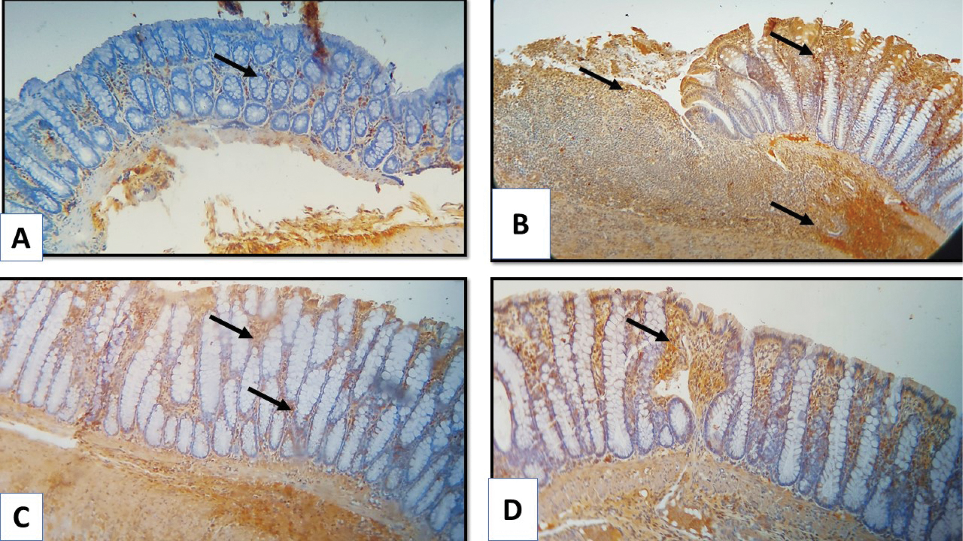
Photomicrographs of colon tissue sections of experimental groups presenting the immunohistochemical expression of IL-1β. A. control group specimen shows a positive cytoplasmic expression of IL-1β in a few cells in the mucosa layer (black arrow); B. untreated AA group specimen shows an increased expression of IL-1β in cells infiltrating mucosa and submucosa (black arrow); C. AA+ sulfasalazine group displays a decreased expression of IL-1β in some cells within the crypts in the mucosa (black arrow); D. AA+ ibudilast group displays a decrease of IL-1β in some cells within the crypts in the mucosa (black arrow). |