|
||
|
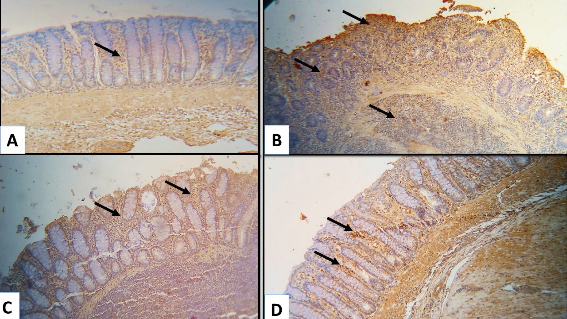
Photomicrographs of colon tissue sections from experimental groups presenting the immunohistochemical expression of TNF-α. A. The control group specimens display a little membranous expression of TNF-α in the in-between the crypts (black arrow); B. The untreated AA group specimens display the increased expression of TNF-α in cells infiltrating mucosa and submucosa (black arrow); C. AA+ sulfasalazine group displays a decreased expression of TNF-α in some cells infiltrating mucosa (black arrow); D. AA+ ibudilast group displays a marked decrease of TNF-α in a few cells infiltrating mucosa (black arrow). |