|
||
|
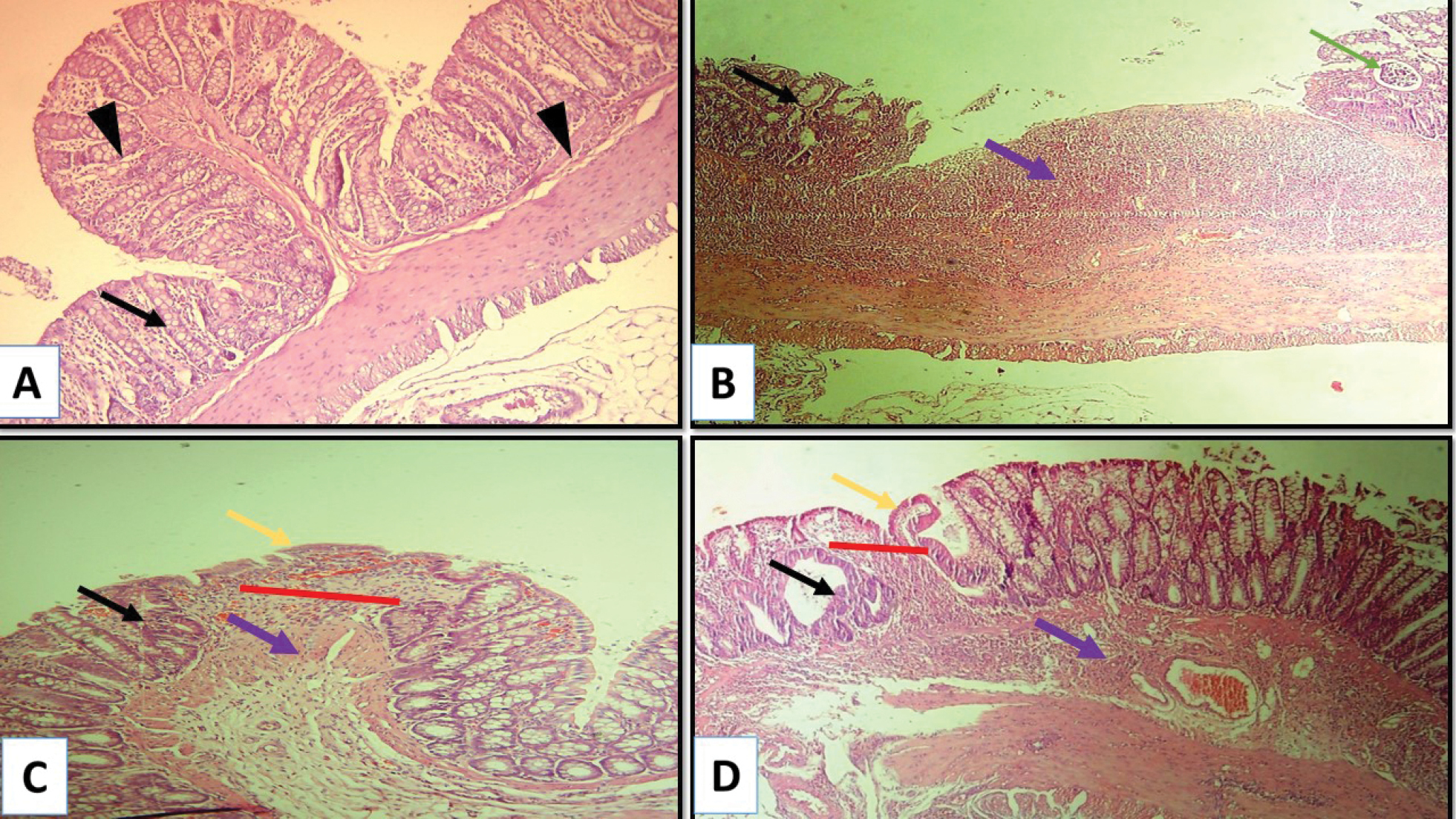
Photomicrographs of colon tissue sections from experimental groups. A. Control group shows intact colonic mucosa and submucosa (arrowhead); B. Untreated AA group shows features of colitis, including extended ulcerated area (black arrow), showing mixed inflammatory cells infiltration and edema (purple arrows), and crypt abscess (green arrow); C. AA + sulfasalazine group shows an improved histological picture with a decreased ulcerated area (red line), less inflammatory cell infiltration (purple arrow) and goblet cells regeneration (black arrow); D. AA + ibudilast group shows improved histologic features with a decreased ulcerated area (red line), a marked decrease in the inter-glandular inflammatory cell infiltration (purple arrow), and goblet cell regeneration (black arrow). H&E stain, X 10. |