|
||
|
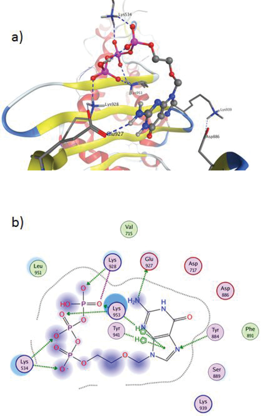
Acyclovir triphosphate and its vicinity in the DNA polymerase pocket after docking procedure: a) 3D plane of view and b) 2D plane of view. The interactions of the ligand in the active site cavity are represented as follows: the proximity contour is depicted with a black dotted line; solvent accessibility, as blue clouds around atoms or blue shadows around amino acid residues; polar amino acids are displayed with pink, while the lipophilic ones are in green. Basic amino acids are outlined with blue and the acidic – with red. Hydrogen bond interactions are depicted with dotted arrows, while the ionic ones are depicted with dotted lines. |