|
||
|
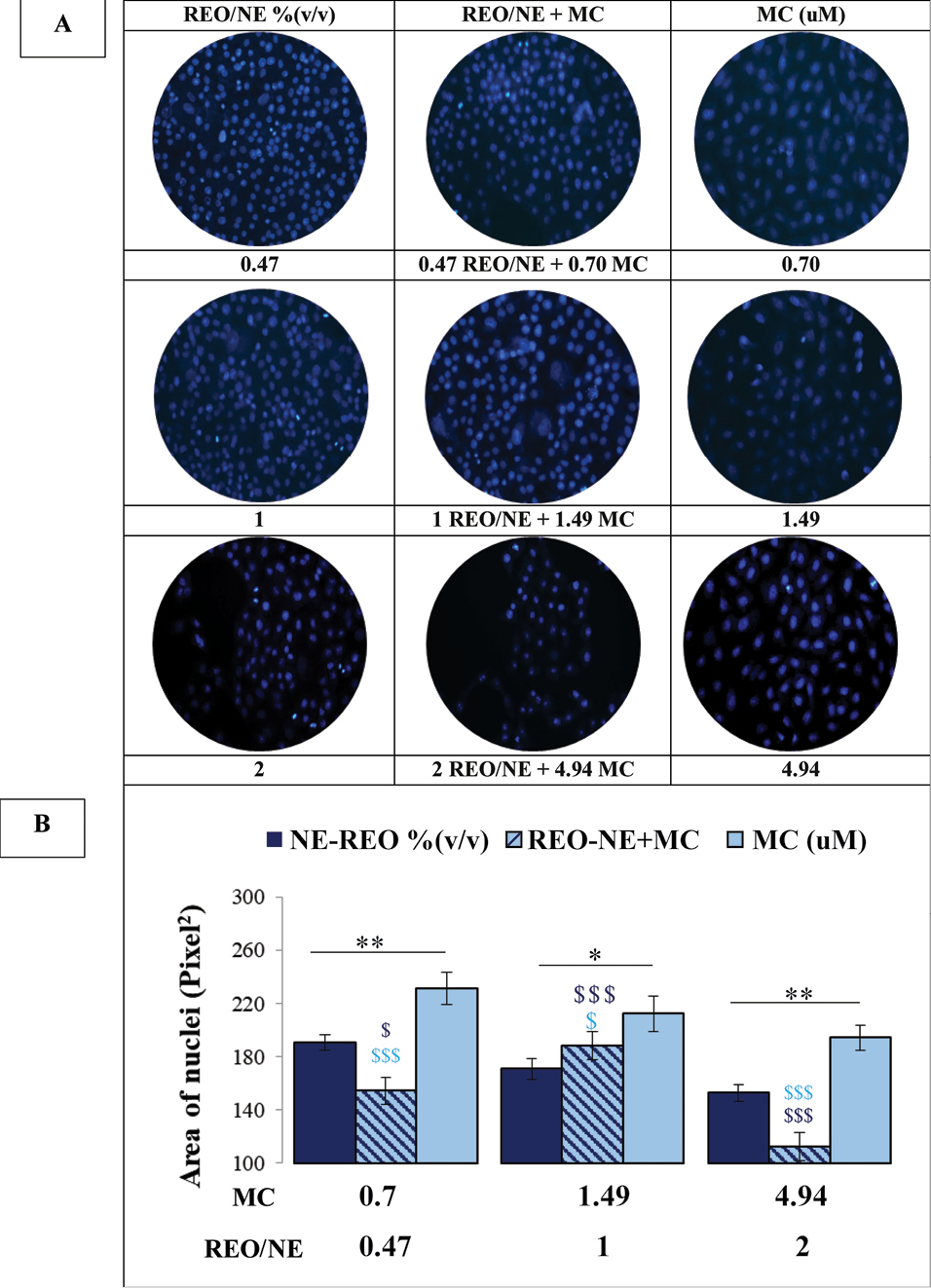
Fluorescence images (x400) of DAPI-stained MCF-7 cells treated with different formulations for 24 h. The treated cancer cells showed abnormalities in the nucleus ($,*p <0.05, **,$$p <0.01, ***,$$$ P <0.001). The symbol * indicates differences between MC and REO/NE; $ indicates differences between REO/NE + MC and each of the individual treatments. Data shown are results from a minimum of three independent experiments. |