|
||
|
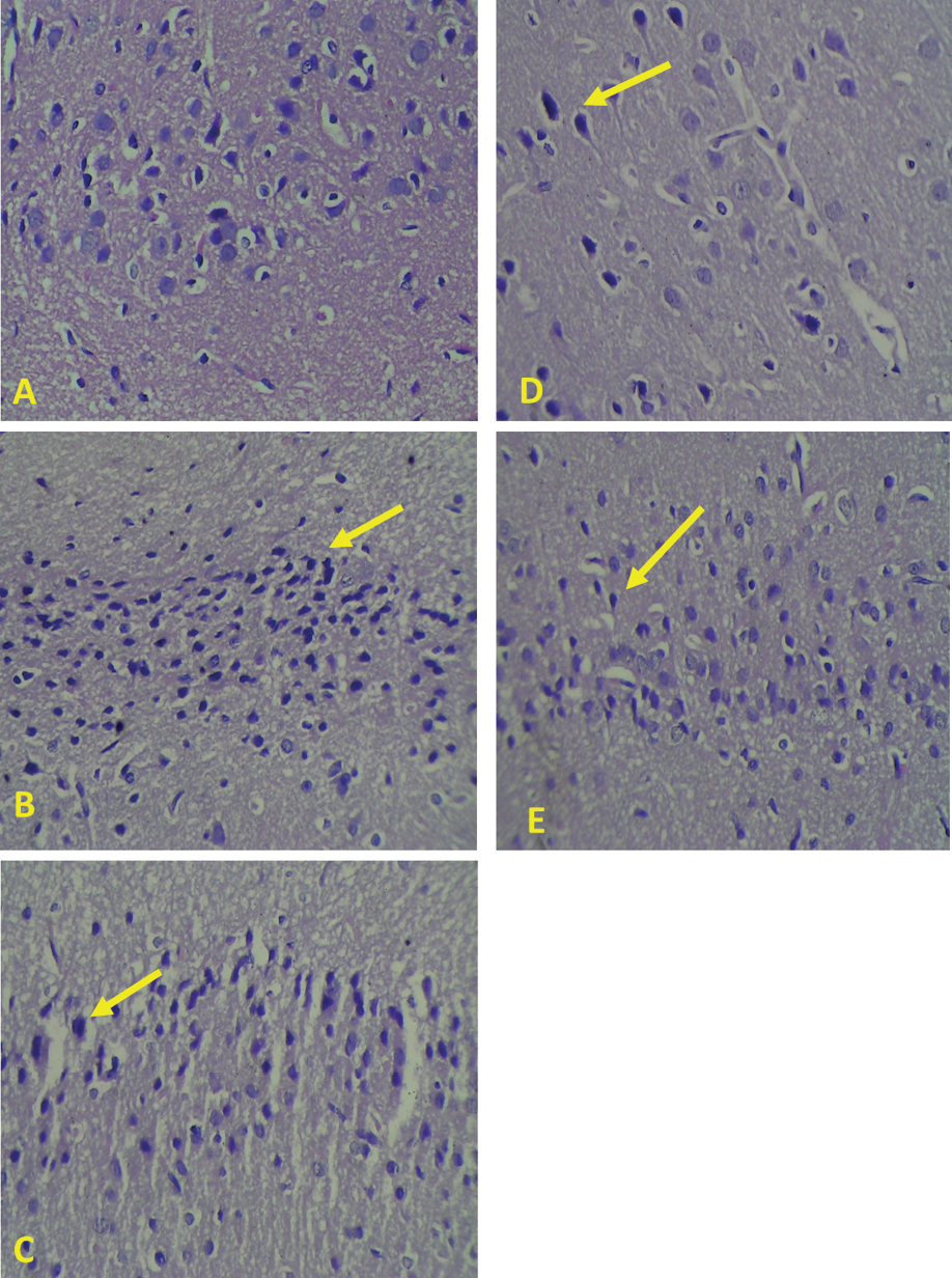
Histopathological picture of brain tissue damage with HE staining, 400× microscope magnification. Description: Histopathological picture of the cerebral cortex area shows degeneration of neuron cells (yellow arrows). A. The normal control group shows a score of 0; B. The negative control group shows a score of 2; C. Group A shows a score of 2; D. Group B shows a score of 1; E. Group C shows a score of 1. |