|
||
|
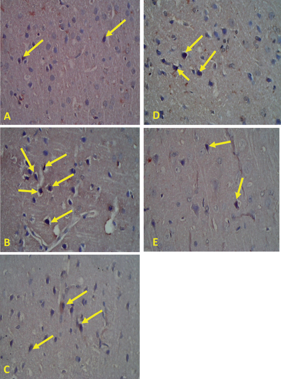
TNF-α immunohistochemical staining with a microscope magnification of 400×. TNF-α expression in the cerebral cortex area shows TNF-α expression in astrocytes (yellow arrows). A. Control group normally expressed 5%; B. Negative control group expressed 15%; C. Group A was depressed 10%; D. Group B expressed 10%; E. Group C expressed 5%. |