|
||
|
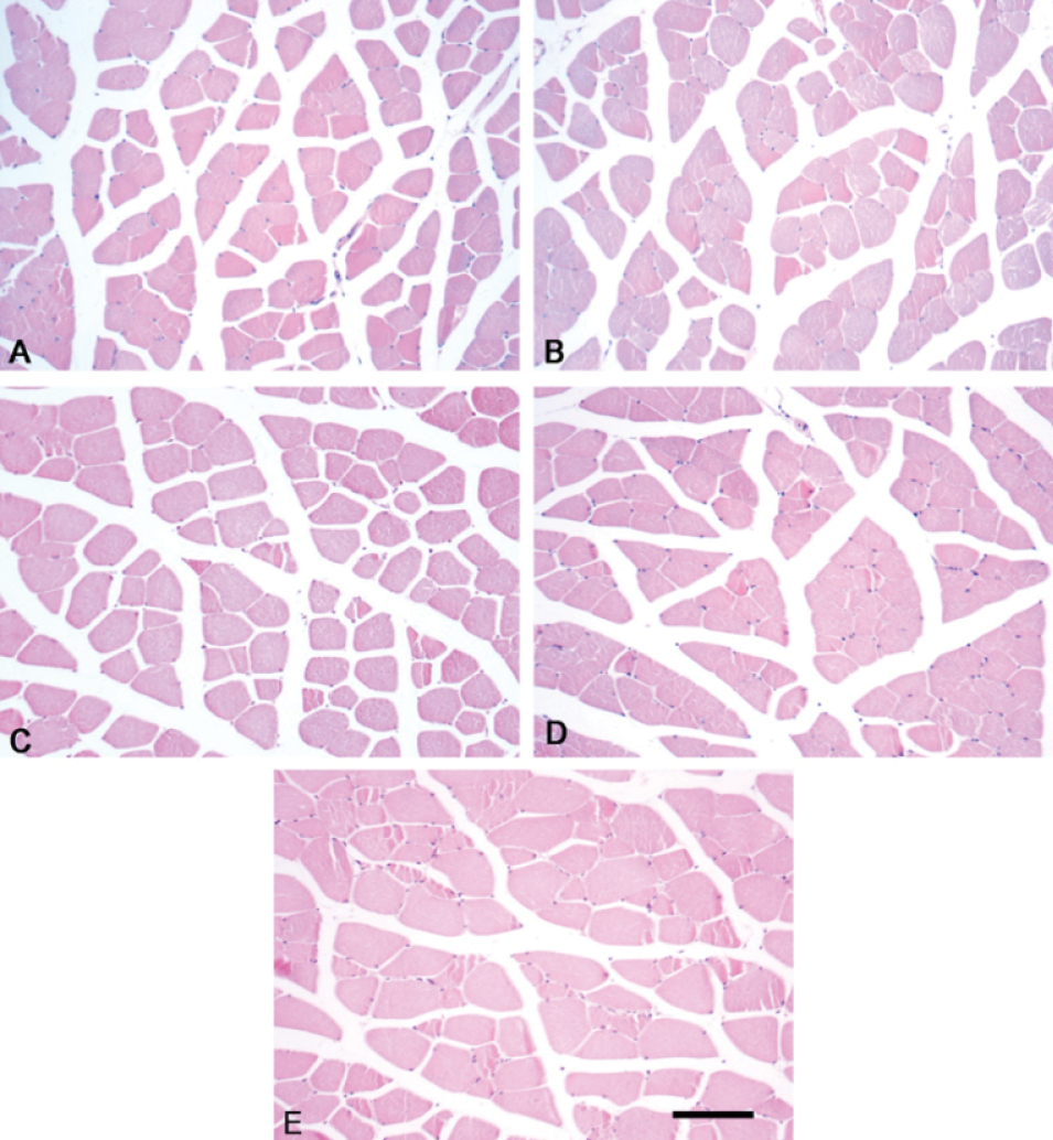
Representative images of hematoxylin and eosin-stained cross sections of the soleus muscle from different groups. Cross-sectioned basophilic nuclei (purple dots) are visible on the periphery of the myofibers. A small number of elongated fibroblastic nuclei between muscle fibers are also visible. (A). Photomicrograph of muscle fibers with regular diameter - control group. (B) Photomicrograph of muscle fibers with moderate diameter - animals treated with CrM 1.5 g/kg/day. (C) Photomicrograph of muscle fibers with larger diameter - animals treated with CrM 3 g/kg/day. (D) Photomicrograph of muscle fibers with moderate diameter - animals treated with CrLys 3 g/kg/day. (E) Photomicrograph of muscle fibers with the largest diameter - animals treated with CrLys 6 g/kg/day. Scale bar: 100 μm. |